Fetal Heart Tone Monitoring Overview
Fetal heart tone monitoring is a critical tool used to assess a baby's status during labor. It can be measured in two primary ways:
- Internal Monitoring: A spiral electrode is inserted through the cervix and attached to the baby's scalp. This method is highly reliable but invasive.
- External Monitoring: An ultrasound transducer is placed on the mother's abdomen. This method is less invasive but can be less reliable due to factors like the baby's position.
Key Components of the Monitor Strip
The monitor strip displays two main lines:
- Top Line: Represents the fetus's heart rate, with a normal range of 110 to 160 beats per minute. The squiggly line shows the trend of the heart rate over time.
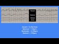
- Bottom Line: Represents the mother's contractions. Each contraction has the following phases:
- Increment: The building or increasing phase of the contraction.
- Acme: The peak of the contraction.
- Decrement: The letting off or decreasing phase of the contraction.
- Relaxation: The period where the uterus returns to its resting state.
Interpreting Fetal Heart Rate Patterns
Accelerations
- Definition: A temporary increase in the fetal heart rate.
- Criteria for a Term Fetus: An increase of greater than 15 beats per minute above the baseline for at least 15 seconds but no more than 2 minutes.
- Common Causes: Fetal movement or contractions.
- Significance: A reassuring sign indicating the baby is healthy, responding appropriately, and receiving proper oxygenation.
- Nursing Interventions: None required. Continue to monitor and document.
Early Decelerations
- Definition: A gradual decrease in the fetal heart rate that mirrors the mother's contraction. The heart rate dips at the start of the contraction and recovers to the baseline by the end of the contraction. The heart rate stays within the normal range (110-160 bpm).
- Cause: Head compression. The contraction compresses the baby's head and the vagus nerve, temporarily slowing the heart rate.
- Significance: A normal, benign finding. For context, this pattern relates to the baby's position in the pelvis, which is further explained in Fetal Station Explained: Understanding Baby's Position in the Pelvis.
- Nursing Interventions: None required. Continue to monitor and document.
Variable Decelerations
- Definition: A sudden, sharp drop in the fetal heart rate (often below 110 bpm) that creates a V-shaped or U-shaped pattern on the monitor strip. The drop is not uniform in shape or timing relative to the contraction.
- Cause: Umbilical cord compression. This reduces oxygen flow to the baby, stressing the fetal heart.
- Significance: A non-reassuring pattern that requires immediate nursing intervention.
- Nursing Interventions:
- Change the mother's position (e.g., knee-to-chest, left lateral, Trendelenburg) to relieve cord compression.
- Administer supplemental oxygen (8-10 liters per minute via mask).
- Stop Pitocin if it is being infused.
- Perform a vaginal exam to check for cord prolapse.
- Notify the midwife or doctor immediately, as urgent intervention may be needed.
Late Decelerations
- Definition: A gradual decrease in the fetal heart rate that begins after the peak (acme) of the mother's contraction and does not recover until well after the contraction has ended.
- Cause: Uteroplacental insufficiency. The placenta is not functioning optimally to deliver adequate oxygen to the baby. For a deeper understanding of placental function, see Understanding Placenta Development and Hormonal Functions.
- Significance: A non-reassuring, ominous pattern that indicates significant fetal distress.
- Nursing Interventions:
- Change the mother's position to improve placental perfusion.
- Administer supplemental oxygen (8-10 liters per minute via mask).
- Stop Pitocin if it is being infused.
- Increase IV fluids to improve placental perfusion.
- Notify the midwife or doctor immediately, as this patient likely requires an emergency cesarean section.
Conclusion
Understanding these fetal heart rate patterns is essential for assessing fetal well-being during labor. Accelerations are reassuring, early decelerations are normal, while variable and late decelerations signal potential problems that require immediate nursing intervention. For a broader review of related maternal assessments, explore Fundal Height Measurement: Maternity Nursing Review for NCLEX.
Hey everyone, it's Nurse Sarah and in this video I'm going to go over fetal heart tone monitoring. And as always,
whenever you get done watching this video, don't forget to access the free quiz. So, let's get started. Fetal heart
tone monitoring is used to help us assess a baby's status during that labor process. And we can measure it various
ways. One way is through internal monitoring. And this is where a spiral electrode is inserted up through the
cervix and put in the baby's scalp. Now, this is really reliable, but it's invasive to the baby and the patient.
Another way is through external monitoring, and this is where they take an ultrasound transducer and put it
across the mother's abdomen. So, with this, it's not going to be as reliable as internal, and it can be affected by
the baby's position, but it's less invasive. Now, whenever you're looking at this fetal heart tone monitoring,
there's a few things you want to be familiar with. First up on the top, typically what you're going to see is
the fetus's heart rate. You have numbers on the side that tells you heart rate ranges. And then you're going to have
this line on this grid that's going to be squiggly. And all it's showing you is how the baby's heart rate is trending. A
normal fetal heart rate is anywhere between 110 to 160 beats per minute. And then below that, you're going to see
another line. And this line is going to represent mom's contractions. So just to review for your maternity exams, let's
talk about the little parts of a contraction. So with this on the monitoring, what you're going to see is
you're going to be able to see the increase of the contraction when it's coming on, hence when it's building.
This is the increment. Then you're going to see the peak of the contraction, the acme. And then you're going to see where
the contraction starts to let off. It hence decreases its decrement. And then you can see the relaxation period. And
this section of the monitoring is going to allow you to see mom's contractions, its frequency, its intensity, and so
forth. So, first up are accelerations. The word acceleration means an increase in rate. And that's what we're talking
about with our baby. This is a temporary increase in the fetus's heart rate. So, for a term fetus, this is a heart rate
greater than 15 beats per minute above its baseline for at least 15 seconds, but no more than 2 minutes. And this can
happen at any time. Typically, what causes it is that the fetus is moving or we have contractions going on. Now, for
this, there's no interventions that are needed. This tells us that our baby is healthy and responding like they should
and they're getting proper oxygenation. Next are early decelerations. And what you want to remember with this is that
the fetal heart rate is going to mirror mom's contractions. So, the baby's heart rate is going to stay within normal
range from 110 to 160. So, you will start to see that the baby's heart rate will dip at the start of mom's
contractions, but it's going to recover at the end of mom's contraction. And here on this screen, you can see you see
baby's heart rate, mom's contraction. They're literally mirroring each other. When mom's contraction starts, that
baby's heart rate dips a little bit, but it's staying within normal. And then at the end of the contraction, that heart
rate recovers nicely. So, what is the cause of this? Well, it's resulting because of head compression. The baby's
head is in that canal. That contraction is happening which is compressing that head. Well, it's also compressing the
vagus nerve which is temporarily causing that heart rate to fall. But once that contraction starts to let up, relieves
that compression, hence that Vegas nerve is not being compressed. So that heart rate recovers nicely. Now with this,
there's no intervention needed. You want to continue to monitor and document. Early decelerations are normal. And then
there's variable decelerations. This is where we have these sudden sharp drops in the fetal heart rate where it's going
less than 110 beats per minute. And then eventually it will try to recover to baseline. So with this, notice on the
bottom we have mom's contractions. When mom's contraction happens, all of a sudden, bam, that heart rate, it drops
down. And it drops so sharply and it tries to increase. And when it tries to increase, it creates this like V or
Ushaped pattern. And that's how I remember this. Remember that variable, we're talking about variable
deceleration starts with V. So whenever this baby's heart rate dropped and so suddenly, it creates like these V- like
shapes. Now, what causes this type of deceleration? Well, it's typically caused by umbilical cord compression. So
whenever mom's having a contraction, it's compressing that umbilical cord. Well, when we compress umbilical cord,
that's literally baby's lifeline. that's going to decrease oxygen. Whenever it does that, that stresses baby's heart
out and hence that heart rate drops. So, this is not a good thing and it requires nursing interventions. So, what you want
to do is you want to change mom's position. There's various positions you can do. You can do knee to chest or you
can do trendelenburgg. This is going to help relieve that compression and help increase oxygen to baby so we don't drop
that heart rate. You also want to give some supplemental oxygen. So about 8 to 10 liters, whatever your protocols say,
via mass to mom so we can get some oxygen to baby. If mom has ptocin running, you want to stop that cuz it
can make this worse. And if need be, you need to perform a vaginal exam to see if you do have maybe cord prolapse and
contact the midwife or doctor because we need immediate intervention. And then lastly, we have late decelerations.
These accelerations are called late because the baby's heart rate is dropping later, hence after the peak of
mom's contractions and doesn't recover until well after that contraction has ended. And here you can see that we have
mom's contraction and then baby's heart rate drops after the peak of that contraction and doesn't recover until
well after that contraction is over. Now, what is causing this type of deceleration? Well, it's typically
caused by uterero placental insufficiency. So, that placenta is not working like it should. It's not
delivering the oxygen that the baby needs. So, this is not good. It requires intervention. Therefore, what you're
going to do, you're going to do well the steps that you did for variable. You're going to change mom's position. You are
going to administer oxygen. You're going to stop ptocin if it's infusing. Also, you may be required to infuse fluids
because this will help provide some profusion to the placenta. and you want to notify that midwife or doctor because
this patient needs immediate intervention and probably needs an emergency C-section. Okay, so that wraps
up this review. And if you're studying for your maternity exams and you'd like some more resources to help you study, I
have a study guide you can get at nurser.com in digital format or you can get this at amazon.com in paperback.
Back.
A normal fetal heart rate baseline ranges from 110 to 160 beats per minute. The top line of the monitor strip tracks this heart rate over time, and any deviations from this range may indicate a need for further assessment.
Accelerations are temporary increases in fetal heart rate that are a reassuring sign of fetal health. They typically occur in response to fetal movement or contractions and show that the baby is well-oxygenated and responsive.
Early decelerations mirror the mother's contraction, with the heart rate dropping and returning to baseline as the contraction starts and ends, and are caused by head compression. Late decelerations, however, begin after the contraction's peak and recover well after it ends, indicating uteroplacental insufficiency and potential fetal distress.
For variable decelerations caused by umbilical cord compression, nurses should change the mother's position (e.g., knee-to-chest or left lateral), administer supplemental oxygen at 8-10 liters per minute, stop any Pitocin infusion, perform a vaginal exam to check for cord prolapse, and notify the healthcare provider promptly.
Late decelerations signal uteroplacental insufficiency, meaning the placenta is not delivering sufficient oxygen to the baby. This pattern indicates significant fetal distress and often requires urgent interventions, including potential emergency cesarean delivery.
For variable or late decelerations, nurses should administer supplemental oxygen at 8 to 10 liters per minute via a non-rebreather mask. This helps improve oxygen delivery to the fetus while other corrective measures are taken.
The bottom line of the strip tracks contractions, showing four phases: increment (building), acme (peak), decrement (decreasing), and relaxation (return to resting tone). This pattern helps correlate fetal heart rate changes with uterine activity.
Keep this summary
Save it to LunaNotes and it becomes a real note in your library — editable, searchable, and ready to turn into flashcards or a diagram. Free to start.
Save to LunaNotesOr summarise for another video.
This summary and transcript were automatically generated using AI with the Free YouTube transcript summary tool by LunaNotes.
Related summaries
Electronic Fetal Monitoring (EFM) Basics: External & Internal Methods Explained
This lecture provides a comprehensive overview of electronic fetal monitoring (EFM), covering external and internal monitoring methods for fetal heart rate and contractions. Learn to interpret key EFM components including baseline, variability, accelerations, decelerations, and contraction patterns like frequency, duration, intensity, and resting tone.

Intrapartum Assessment & EFM Interpretation: Focused Nursing Guide
This mini-lecture covers the five essential focused assessments for intrapartum patients: pain, contractions, leaking of fluid, vaginal bleeding, and fetal movement. Learn how to subjectively and objectively evaluate each component, including a practical palpation trick to gauge contraction intensity, and understand the critical importance of fetal movement as an indicator of fetal oxygenation.

Intrauterine Resuscitation: Fetal Heart Rate Categories & POISON Mnemonic
This lecture breaks down how to interpret fetal heart rate categories (I, II, III) and apply the POISON mnemonic for intrauterine resuscitation. Learn when to intervene, including position changes, oxygen, IV fluids, and turning off oxytocin, with real clinical examples to build critical thinking skills.

Intrapartum Nursing: Labor Stages, EFM Interpretation, and OB Emergencies
This comprehensive lecture summary covers essential intrapartum nursing concepts including true vs. false labor assessment, the four stages of labor with their phases, fetal heart rate monitoring interpretation (baseline, variability, accelerations/ decelerations), and critical obstetric emergencies like cord prolapse, shoulder dystocia, and amniotic fluid embolism. A must-review for nursing exams and clinical practice.

Stages of Labor, Cervical Change & True vs False Labor Explained
Understand how to differentiate true labor from false labor and learn the four stages of labor. This comprehensive guide covers key assessments like dilation, effacement, and station, plus signs of approaching labor for nursing students and healthcare professionals.
Most viewed summaries

A Comprehensive Guide to Using Stable Diffusion Forge UI
Explore the Stable Diffusion Forge UI, customizable settings, models, and more to enhance your image generation experience.

Kolonyalismo at Imperyalismo: Ang Kasaysayan ng Pagsakop sa Pilipinas
Tuklasin ang kasaysayan ng kolonyalismo at imperyalismo sa Pilipinas sa pamamagitan ni Ferdinand Magellan.

Mastering Inpainting with Stable Diffusion: Fix Mistakes and Enhance Your Images
Learn to fix mistakes and enhance images with Stable Diffusion's inpainting features effectively.

Pamamaraan at Patakarang Kolonyal ng mga Espanyol sa Pilipinas
Tuklasin ang mga pamamaraan at patakaran ng mga Espanyol sa Pilipinas, at ang epekto nito sa mga Pilipino.

How to Install and Configure Forge: A New Stable Diffusion Web UI
Learn to install and configure the new Forge web UI for Stable Diffusion, with tips on models and settings.
Found this summary useful?
Take it with you. One click puts it in your own LunaNotes library.
Save to LunaNotes