Intrapartum Nursing: High Points Synopsis
Defining True Labor
- Two essential components:
- Progressive contractions (increasing in intensity, frequency, and duration)
- Cervical change (dilation, effacement, or fetal descent/drop in station)
- Not all three changes needed, but some cervical change must occur from progressive contractions
For a deeper breakdown of how to differentiate true labor from false labor and assess cervical change, see our guide on Stages of Labor, Cervical Change & True vs False Labor Explained.
Sterile Vaginal Exam (Cervical Assessment) - Three Assessments
- Dilation: How open the cervix is (measured in cm, from closed to 10 cm)
- Effacement: How thinned the cervix becomes (measured in percentage, from thick to 100%)
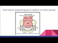
- Station: Fetal presenting part's relation to maternal ischial spines
- Negative numbers: Baby still high in pelvis
- Zero station: Presenting part even with ischial spines
- Positive numbers: Baby descending for delivery; +2 to +3 typically shows crowning externally
The Four Stages of Labor
- Stage 1 (Dilation): From onset of contractions/cervical change to complete dilation (10 cm)
- Stage 2 (Pushing): From complete dilation to delivery of baby
- Stage 3 (Placental Delivery): Ideally within 30 minutes of baby's birth
- Stage 4 (Postpartum Recovery): Immediate postpartum period for mother
Stages 1 & 2: Phases
Stage 1:
- Latent Phase (up to 5 cm dilated):
- Contractions irregular, less intense
- Patient conversational between contractions
- Best time for patient education and task completion
- Active Phase (6 cm or more):
- Contractions closer, stronger, more regular
- Transition Phase (8-10 cm): Often accompanied by nausea/vomiting, "I can't do this" statements, turning inward
Stage 2:
- Latent Phase: Fully dilated but not actively pushing (may lack urge, especially with epidural)
- Active Phase: Fully dilated and actively pushing
- Note: Patients without epidural rarely have a Stage 2 latent phase
Labor Assessment Framework (Intrapartum 5)
- Vital signs (critical baseline)
- Pain (subjective, use 0-10 scale, note location/descriptors)
- Contractions (subjective + objective measurement)
- Leaking of fluid (note time of rupture, fluid color)
- Vaginal bleeding vs. bloody show
- Bloody show: Red-tinged mucus, common with dilation/effacement/exams
- Bright red bleeding: NOT assumed normal, requires investigation
- Fetal movement
Fetal Heart Rate (EFM) Interpretation
Baseline: Average FHR rounded to nearest 5 (ends in 0 or 5); normal range 110-160 bpm
Variability (4 categories):
- Marked: Likely can't determine baseline
- Moderate (6-25 bpm): Desired category
- Minimal (1-5 bpm): Less than 5 but not absent
- Absent (0 bpm): Flat line, no beat-to-beat changes
For a detailed overview of monitoring methods and their indications, review our guide on Electronic Fetal Monitoring (EFM) Basics: External & Internal Methods Explained.
Accelerations: FHR speeds up; 15 beats above baseline, lasting at least 15 seconds. Indicates good oxygenation.
Decelerations: FHR slows down. For a more detailed explanation of each deceleration pattern, see Fetal Heart Tone Monitoring: Accelerations & Decelerations Explained. | Type | Cause | Shape | Key Feature | |------|-------|-------|-------------| | Variable | Cord compression | Sharp V-shaped | Abrupt onset/return, often changes with position | | Early | Head compression | Flat U-shaped | Mirrors contraction (deepest point matches contraction peak). No intervention needed. | | Late | Placental insufficiency | Flat U-shaped | Begins after contraction starts; returns to baseline after contraction ends. Always warrants intervention. |
Contraction Assessment
- Frequency: Start of one contraction to start of next (range in minutes)
- Duration: Start to end of each contraction (range in seconds)
- Intensity & Resting Tone: Require palpation at contraction peak; abdomen should be soft between contractions
Intrauterine Resuscitation Interventions
- Variable decelerations (cord compression): Change maternal position (easiest/non-invasive)
- Late decelerations (placental insufficiency):
- Turn off Pitocin if running
- Administer oxygen
- Increase IV fluids
- Change maternal position
- Minimal/absent variability:
- Can't fix CNS depression externally, but can improve oxygenation
- Administer oxygen to increase variability
- Fetal tachycardia + moderate variability: Oxygen NOT warranted; usually caused by maternal fever. Treat infection to lower fever/baseline.
For a concise, step-by-step approach to these interventions, review the Intrauterine Resuscitation: Fetal Heart Rate Categories & POISON Mnemonic guide.
Obstetric Emergencies
Umbilical Cord Prolapse
- Signs/Symptoms: Abrupt, severe variable deceleration → prolonged or fetal bradycardia
- Interventions:
- Elevate presenting part (hand)
- Turn off Pitocin
- Bolus IV fluids, give oxygen
- Position mother in Trendelenburg or hands-and-knees
- Proceed for emergency Cesarean (ideally within 10 minutes)
Shoulder Dystocia
- Key Timeline: 5 minutes from head delivery to delivery to prevent poor outcomes (hypoxia, ischemia, brain damage)
- Interventions:
- McRoberts maneuver: Flex maternal legs up to ears (opens pelvic outlet)
- Suprapubic pressure: Pressure above pubic bone to dislodge shoulder
- If persistent: Flip mom to hands-and-knees; internal maneuvers by physician
Non-Reassuring Fetal Heart Tones
- Category 3 strips always warrant delivery if can't correct immediately
- Concerning Category 2 strips + failed interventions = likely emergent delivery
Amniotic Fluid Embolism (AFE)
- Mechanism: Amniotic fluid enters maternal circulation; body's severe reaction (like anaphylaxis) → cardiopulmonary collapse (NOT a blockage like PE)
- Signs/Symptoms:
- Sudden shortness of breath, chest pain
- Overwhelming sense of doom
- Cardiac and respiratory arrest
- Management:
- Full code blue, CPR, intubation
- Blood product transfusion, ICU admission
- If during Stage 2: Expedite delivery
Uterine Rupture
- Risk Factor: Previous uterine scar (most commonly Cesarean; also myomectomy)
- Signs/Symptoms:
- Abrupt cessation of contractions
- Severe, abrupt FHR change → bradycardia
- Maternal vital sign changes (↓BP, ↑HR, ↑RR, ↓O2 sat)
- Vaginal bleeding; may palpate fetal parts abdominally
- Management: Emergency delivery + stabilization
Intrapartum Hemorrhage (from abrupted placenta or vasa previa)
- Same emergency management: Emergency Cesarean, stabilization, bleeding control
Urgent (Non-Emergent) Situations
Cephalopelvic Disproportion (CPD)
- Baby's head too large for maternal pelvis
- Often detected by failure of fetal descent to zero station
- Warrants Cesarean delivery (not necessarily emergent; can be "as soon as possible")
Intra-Amniotic Infection (Chorioamnionitis)
- Infection in uterine cavity during labor
- Requires antibiotics, fluid resuscitation
- Mom febrile; baby likely tachycardic
- Goal: Deliver baby sooner rather than later
Prolonged Labor
- Cause depends on prolonged first vs. second stage
- Determines possible interventions
Meconium-Stained Fluid
- Increases risk for aspiration, pneumonia, mechanical obstruction after delivery
- Notify provider and NICU/special care team
- May indicate fetal distress (stress/hypoxia) that caused meconium passage
- Meconium itself usually doesn't actively harm fetus in utero
Operative Vaginal Deliveries
- Vacuum: Maximum 3 pulls ("3 strikes, you're out"); may leave marks on fetal head; requires informed consent
- Forceps: More invasive, higher risks for maternal/fetal tissue damage, lacerations, bleeding
- Both require: Informed consent with risk/benefit discussion, appropriate indications, and post-delivery monitoring for complications
For a focused review of all intrapartum assessment components including EFM interpretation, see the Intrapartum Assessment & EFM Interpretation: Focused Nursing Guide.
Okay, so last mini lecture for intrapartum content. This is going to be a synopsis of high points we'll call it.
So not that this is new content or not that this is um necessarily all-incclusive of content that uh we
need to understand for the exam, but we're going to hit on the high points of the larger picture pieces that we've
discussed and applied in class. So to begin inch part we're talking about labor. So how do we define true labor?
Remember that is um two parts we have to have contractions that are progressive in nature. So um increasing in
intensity, increasing in frequency, increasing in duration that are causing cervical change. So we have dilation or
aacement um or drop down of station for the fetus um in the birth canal. And it doesn't need to be all of those things,
but we have to have some kind of change occurring to the cervix as a result of progressive contractions to deem a
patient in labor. And then when we look at what three assessments are included in a sterile vaginal exam or cervical
assessment, we know that we are looking at dilation, easement, and station. So dilation being how open the cervix is.
Remember, we measure that in centimeters, and we're going from closed to 10. Easement is how thick and or thin
that the cervix gets throughout the labor progress. Um, and remember that one's measured in percentage. So, we're
going from thick all the way to 100% of where there's no um no width or length left of the cervix when we're ready to
deliver when we're fully completely dilated. And then fetal station being fetal uh the fetus's presenting part
ideally for vaginal delivery is ahead, but whatever presenting part we have. And then the relation of that presenting
part to the isial spines of the pelvis for the laboring woman. And we know that we have negative numbers, which means
baby's still really up high in the pelvis. Zero, meaning baby's presenting part is even with the issial spines,
essentially closing off the exit to anything except baby. And then positive numbers, meaning baby is on the way out
for delivery past just engagement. Um, in that we usually get to about plus 2 to + 3 is when we see crowning visually
from external. And then our four stages of labor. So we know we have stage one which is the
labor process itself, the dilation part. So we're from the beginning of contractions and cervical change
occurring all the way through to where we completely dilate. Um and that is our starting point for stage two. Stage two
is the pushing or the um waiting to push if we're laboring down and we're in that latent phase. Um stage where we are
starting with full dilation and ending with delivery of baby. Stage three is our delivery of the placenta. Ideally
happens within less than 30 minutes. And then stage four is the postpartum recovery period for mom. So those are
our four stages. And then we know that our first and second stage have phases. So the first phase of the first stage is
our latent phase. That is all the way up to 5 cm dilated. Contractions are usually not super super close together.
Um they might be irregular in both frequency and duration. There is some type of of cervical change happening and
the patient during the latent phase of stage one is usually still able to talk and hold conversation especially in
between contractions. They are comfortable or at least not severely in pain breathing through contractions when
they're occurring. This is the time that they are most receptive to information, to education, to um looking ahead as to
what what to expect next. That is the time where we try to get all of those those tasks and um objects completed.
And then stage one phase two is our active labor. So 6 cm or more in dilation usually much more aacement um
contractions are closer together, stronger, lasting longer, more rhythmic in nature or more regular in nature. Um
and then we have that to you know from 6 centimeters on remembering that our patients who do not have an epidural or
any type of anesthesia. Um often also experience transition phase which is in that 8 to 10 cm right towards the end.
Um and a lot of times that is accompanied by nausea and vomiting um turning inward. I can't do this. I want
to go home. those kind of statements um usually means it's about to they're about to um the first and second phase
of stage two is just the difference of if we are fully dilated and pushing which would be our active phase or we're
fully dilated and we're waiting to push because we don't have the urge or the epidural is blocking that urge and
there's no urgency to do that. Um, also remembering that if a patient does not have an epidural stage 2 latent
phase really probably not going to happen much for them. So our labor assessment remember we said
we have we're always going to start with vital signs are a are a critical component of any any patient assessment.
So we need to um obtain vital signs and we need to know what our normal ranges is are for vital signs. we are going to
ask the patient about pain. Um and then along with that knowing that what we are doing for pain andor support during the
labor process will vary patient to patient. Um we need to know what is what is acceptable methods of pain control
both non-farmacologic and pharmacologic. Um we need to know the difference between anesthesia and analesia. Right?
So we have our epidural spinal or general anesthesia and then we have our analesics pain control. um via other
medications that are mostly systemic in nature. Our intraartum five, so we talked a little bit about that pain
piece. And then we have our contractions, leaking, bleeding, and fetal movement. So remembering that pain
is both subjective. We're asking the patient and then we need to follow up with descriptors, location, um what kind
of pain, a 0 to 10 scale for pain, um because that pain might not always be related to labor pain itself. um our
contractions, subjective and objective measurement. Same for leaking of fluid and vaginal bleeding. If our patient has
had rupture of membranes, we need to know the time that it occurred and what color the fluid was. Um if the patient
is experiencing vaginal bleeding, that is never to be assumed normal, but it also doesn't mean that something is
catastrophically wrong. Um remembering that bloody show and vaginal bleeding are
different things. Bloody show is more of a red tingy mucus um that is common as the cervix dilates and a faces and
especially with um manipulation from vaginal exams that increases bloody show. Um vaginal bleeding bright red
bleeding is not to be assumed normal and needs further investigation. Then we have our EFM and contraction
interpretation and remembering what we what the different categories are we look at for both of those and then what
we do or intervene upon based on our findings. So for our fetal heart rate we know we are starting with our baseline
first and foremost always average essentially of our fetal heart rate rounded to the an increment of five. So
ending in a zero or five we would like that baseline to be between 110 and 160 cuz that's our normal range. After we
determine baby's baseline, we look at variability, remembering that we have four categories of variability. Um,
marked variability means we probably couldn't determine a baseline to begin with. So, that would be um an easy
checks and balance system for us. And then the other three categories of variability being moderate, minimal, or
absent. Absent meaning none at all, flat line, no jagged, no um no beat to beat changes. We don't get that back and
forth. Um minimal being less than five beats, but so one to five, right? because if it was zero it would be
absent. 1 to 5 for minimal and then 6 to 25 being our moderate which is the um category of variability we would like to
see accelerations meaning fetal heart rate speeds up. We know we're looking for 15
by 15. So we want 15 beats higher than baseline and we want that um acceleration to last at least 15
seconds. So from the time the fetal heart rate leaves baseline until the time it returns to baseline should be at
least 15 seconds. And that would be considered an acceleration. Accelerations caused by oxygenation. We
love an oxygenated fetus. So we mark that those are there. We are reassured and we continue on. Decelerations
meaning the exact opposite. So instead of accelerating, we're decelerating. The fetal heart rate is slowing down. And we
know that we have various different kinds of decelerations. And the type of deceleration tells us what the cause of
the deceleration is. So variable decelerations caused by chord compression. The ones that look like
these, very sharp, abrupt to decrease, very um quick return to baseline. Our early decelerations are caused by head
compression. So our earies and our late, remember, often the same shape or look very similar in shape. They're flat U
shapes instead of that variable sharp V. Um our early decelerations caused by head compression. Those decelerations
are going to mirror the contractions. So the the deepest point of the deceleration is going to match up with
the highest point of the contraction and those are caused by head compression and we don't have to do anything about
them except document and continue on. Late decelerations look again the same shape as our earies. However, they uh
deceleration begins after the contraction has started and the baby's baseline or the baby's heart rate does
not return to baseline until well after the contraction is over. It comes back to baseline late. That's how we
determine it's a late deceleration. Late decelerations are caused by placental insufficiency and should always always
warrant intervention of some sort. Our contractions, we are looking at the frequency of duration or frequency of
contractions, meaning start of one contraction to the start of the next. Duration being the start to end of each
contraction. And both of those things can be a range. They're not um always the exact same when we're looking at a
strip. So a range is fine. For frequency, that's usually a range of minutes. And for duration, that is more
frequently a range of seconds. Intensity and resting tone. Um, you note that we have a little asterk by those. Those
those are going to require palpation to objectively assess if we are using external monitoring, which we mostly do.
So, remember that the um the nurse needs to palpate during the peak of a contraction for intensity and the um
resting tone would then be between contractions. We hope to feel that the abdomen is soft. That means everything
is relaxed and there's adequate blood flow and oxygenation going to the fetus in between.
So we talked um through intrauterine resuscitation in class and we looked at some strips and we kind of applied which
pieces um or which interventions within uh intrauterine resuscitation are warranted based on fetal changes and
what those changes are. Um so ones we know again not all-incclusive but we know that if we have variable
decelerations uh that's cord compression our easiest and most non-invasive intervention for that is to change mom's
position change the position we change the pressure on the cord we can alleviate those variable dels um if we
have late decelerations uh that indicates the baby is not tolerating those contractions well if we have
ptocin it needs turned off um if mom is um not well oxygenated we can give extra oxy oxygen for that we can um increase
IV fluids, change her position, try to improve the profusion and or oxygenation of the blood flow to the uterus which to
the placenta to baby. Um if we have minimal or absent variability, we can't do a lot about um
depression of the central nervous system from the outside aside from not giving mom meds that would do that. But we can
influence oxygenation. So we can give oxygenation for minimal or absent variability. Hyper oxygenate mom
theoretically which pushes more oxygen to the fetus and then improves our variability. So those are some examples
that we talked about. Um remembering that we always want to be able to identify what the rationale is for any
intervention. Um and that will help you decide based on the change that you're seeing what the appropriate intervention
is. Um for example if we have fetal tacoc cardia um but we have moderate variability no d
cells no axels but we have fetal tacic cardia um giving mom oxygen is not warranted because we have moderate
variability and oxygen is not going to change the fetus's baseline. Therefore that's an unnecessary intervention.
Right. Um, we said that fetal tacic cardia is most often, if not always, caused by maternal fever. So, what we're
going to do in regards to maternal fever is we're going to give her medication to treat whatever is causing her fever,
which is more than likely an infection. We're going to treat the infection. If we bring down her fever, we decrease her
body temperature. Baby's baseline will then decrease as a result. And then we have a normal range baseline.
We talk about our complications. We have we know we have some that are concerning and then some that are emergent. Right?
So these are more our emergent scenarios. Um an umbilical cord prolapse signs and symptoms of that would be an
abrupt severe variable deceleration that is either extensively long and or results in fetal brada cardia because
the compression of that cord is not being relieved if we have a true cord prolapse. Um what we do about that we
elevate whatever the presenting part is. We turn off the toin if it's running. We bololis fluids. We give oxygen. We
position mom um in a trendelenberg position or on their hands and knees to try to help gravity pull the weight and
pressure from the fetus off of the umbilical cord. We use our hand, elevate that presenting part off the cord to
decrease that pressure and then we proceed for an emergency C-section. Um we hopefully by the time we identify
that there is a cord prolapse we are in the we are in the O delivering that baby within like 10 minutes or less would be
ideal for a shoulder dystocia when baby's shoulder gets stuck on the way out. Um so we cannot diagnose the
shoulder dystocia until after the head is delivered and then the shoulder gets stuck and we have specific interventions
that we um need to apply for that. Mc Roberts is a very um initial intervention that's when we have
maternal flexion of the legs. So essentially pushing her legs all the way up to her ears as as far as it can go.
The rationale for that is that it opens the pelvic outlet as much as it can possibly be to hopefully relieve um the
stuck shoulder. Uh in addition to that super pubic pressure, so pressure of the nurse usually right above the pubic bone
of mom, pushing ideally on baby's shoulder to pop it underneath that pubic bone. Um that's going to help dislodge.
Um if those things don't work, we can flip mom on her hands and knees. There are internal maneuvers that the
physician can do to try to get baby unstuck. Um, some of them more invasive as we go along, but we know that we have
a 5minute kind of time limit from the time baby's head's delivered and we diagnose the shoulder dysia to get baby
out without really really poor outcomes related to hypoxia and tissue eskeeia and irreversible
brain damage. Um, other emergent situations non-reassuring fetal heart tones. So always are category 3es,
right? anything that warrants a category 3 strip warrants a like if we can't fix it right now we need to deliver and then
I would also add to that that we have if we have a concerning category 2 strip that we are still in category 2 but we
have done all of the interventions we can andor know how to do um and we're not seeing an improvement that that
fetal heart rate is non-responsive um that is probably going to warrant an emerent situation
so Then we look at amniotic fluid embolism which we know is one of the um most severe medical emergencies
in OB that is um the mortality rate of which is extremely high for mom and also for subsequently then baby if it is um
if it occurs prior to delivery. So we think of amniotic fluid embolism sort of in the way that we we think of a
pulmonary embolism, but it isn't the same reaction. So it's an embolism in the fact that there is a small amount of
amniotic fluid that is pushed into the circulatory system during labor and or delivery or shortly after. Uh however,
it is not an embolism that it causes a blockage of some sort to some part of the body that then causes the squellle
afterwards. It is the body's reaction to the amniotic fluid in the circulatory system. Um, which is almost like a
it sees it as a foreign object almost to the point of like an anaphylactic allergic reaction. However, it's not
it's not the same as that. Um, that's just the most the closest thing I think we can um liken it to. But it is a
severe reaction of the body to the foreign substance of which is the amniotic fluid in the circulatory system
that causes a cardopulmonary collapse. Um so if our patient is exper experiencing an amniotic fluid and
embolism, the signs and symptoms are going to somewhat fall in line with what we would expect
for a pulmonary embism is that they they feel like they have shortness of breath or they're having trouble breathing or
they might experience chest pain. They have an overwhelming sense of doom. Um that something is not right even if they
cannot verbalize what is not right or what is wrong. Uh and then they collapse. They uh their heart they will
go into cardiac and respiratory arrest. Um it is very sudden. It is very severe. It will require CPR. It will require a
full code. It will often require um intubation and transfusion of blood products and an admission a lengthy
admission to an ICU um to support the patient's recovery. If this occurs prior to delivery during the labor process,
usually stage two, um it is expediently required that we deliver the fetus as quickly as possible. Um and then we need
to alert all of the people. This is the full code blue situation. You want as many hands on deck as possible. Um, so
you realizing that CPR and quick notification to all of the interdisiplinary personnel that we need
to take care of this patient are our top priorities. Um, uterine rupture most likely um
occurring for a patient who has had a previous uh incision and scar on their uterus. Most likely that's a cacaian.
doesn't have to be. They could have had a momectomy or some other type of um surgical procedure on the uterus which
of which leaves a scar on the muscle. Um but most frequently it's a previous cacaian um whether they're trying to
vacc um or that we just got really careless with the use of arptocin and we weren't
monitoring appropriately or whatever that might be. Uh we see a an abrupt cease in contraction activity, uterine
contraction activity. We see a severe and abrupt change to the fetal heart rate that usually ends up in brada
cardia. We see changes in mom's vital signs that indicate an intraartum hemorrhage of some sort. Uh i.e. blood
volume leading the circulatory system. So our decreased blood pressure, increased pulse, increased respiratory
rate, decreased O2 saturation. Um the mom may or may not stay conscious for an extended period of time
depending on how severe the abruption is and how much blood loss occurs very quickly. Um but we will be moving
towards delivery expediently. Um there is usually a good deal of vaginal bleeding associated with this. Uh a lot
of times you can palpate the fetal parts through the abdomen because they're no longer in the uterus. Um but this is
definitely one of the emergency situations in which an emergency delivery and then stabilization is
required. Um and then addition to that the intrapartum hemorrhage that can uh stem from an abrupted placenta where the
placenta separates from the uterine wall prematurely or from the rupture of a vasa previous situation. Um again all
treated very similar emerent cesarian delivery stabilization um and control of bleeding and support
for recovery. So then we have our non-emerent which is urgent situations um in which
we are not in an emergency situation. However, uh there are many interventions needed. Some of them more urgent
intervention. Um and we want to monitor the progress of labor for these different complications.
So, see pelvic disproportioning that that essentially means baby's head is not conducive for mom's pelvic size and
therefore won't be exiting the vaginal route. Uh we often in we often pick up that this could be an issue when we have
failure of the fetus to descend into the pelvis. uh failure to get to that zero station. And this warrants um a
C-section delivery. Not necessarily that it needs to be emergent. Baby's tolerating everything well and still
looks reassuring on the monitor. It can be, you know, as soon as possible kind of thing, but it doesn't need to be a
general anesthesia emergency situation. Um II intramniotic infection or coro. Um coro is the old term, II is a new term,
but essentially means an intrauterine infection. So, an infection has um been diagnosed in the uterine cavity while
baby is still on the inside during labor. Uh this is going to require antibiotic therapy, fluid resuscitation
as far as like replenishing fluid. Mom usually has a fever. Uh baby will likely be tacocartic at some point during
labor. Um we might see an elevated white count. We might test the fluid. All of the things that go along with infection.
Um but we want baby delivered sooner rather than later in that situation. prolonged labor. So again, depending on
if that's prolonged first stage or prolonged second stage tells us what the cause or underlying um underlying
rational for that might be and then that often tells us what if there's anything we can do to fix it.
Mcconium stain fluid baby passing mononium while still in uterero um we might we always know note that it's
there. Um the provider needs to know um NICU or special care whoever might be helping with baby resuscitation after
delivery needs to know. um it increases the risk for aspiration and pneumonia and mechanical obstruction for breathing
for baby after delivery. It's not so much something that we need to um it's not going to impact
really severely usually um the actual labor process. We usually start to see baby have issues after delivery with
this. Although we could have um the theory is that babies passonium in uterero because of a stressful invent
event or environment or hypoxia something like that. So we might see changes on the fetal monitor that
indicate the fetus is distressed that led to the passing of mononium. It's not usually that the mcconium is actively
causing an issue in the fetus while they're in utero. Um and then operaginal deliveries. So, if we're using a vacuum,
remembering we get three pulls, three strikes are out, um that we might see some marks on the fetal head afterwards,
that we need to educate mom about what it sounds like if it pops off, how many times we'll try, what is normal,
abnormal, um watching the fetal heart rate strip, when is it indicated that we can do a vacuum delivery, um what
findings do we need to be able to see before we would try that. Same thing for forceps. forceps being even more um
invasive and problematic as far as risks associated to both mom and baby for tissue damage and lacerations and
bleeding um and all of those pieces. So again, all of these should include informed consent to the patient prior to
applying any of those that there should be a full risk benefits discussion on what the risks are um and then what
happens if we don't do this as evidenced by you know what what the situation calls for. Um so all of the that should
should be discussed prior to and then knowing the risk that it increase when we use either of those um to look for in
mom and or baby after delivery and that is our is our little synopsis for interpartum. So again it's not in
all inclusive that is not everything we could need to know for the exam but that is um a quick synopsis of all the high
points for the different various pieces that we're looking at. So if we understand that well we can apply that
knowledge to all of the different um pieces that we are going to be looking at at on the exam and in clinical and in
simulation.
True labor requires two components: progressive contractions that increase in intensity, frequency, and duration, and some degree of cervical change (dilation, effacement, or fetal descent in station). Not all three cervical changes must occur, but at least one must be present alongside progressive contractions. This is assessed clinically through monitoring contraction patterns and performing sterile vaginal exams to evaluate cervical dilation, effacement, and station.
FHR baseline is the average rate rounded to the nearest 5 (normal 110-160 bpm). Variability is categorized as moderate (6-25 bpm, desired), minimal (1-5 bpm), absent (0 bpm), or marked. Accelerations (15 beats above baseline for 15 seconds) indicate good oxygenation. Decelerations are categorized as variable (cord compression, sharp V-shaped), early (head compression, mirrors contraction), or late (placental insufficiency, begins after contraction and returns after it ends, requiring intervention). Late decelerations always warrant immediate action.
For variable decelerations (cord compression), change maternal position. For late decelerations (placental insufficiency), turn off Pitocin, administer oxygen, increase IV fluids, and change position. For minimal/absent variability, improve oxygenation with oxygen; for fetal tachycardia with moderate variability, treat maternal fever with antibiotics (oxygen is not needed). The POISON mnemonic (Position, Oxygen, IV fluids, Stop Pitocin, Notify provider) guides these steps, escalating to emergency delivery if interventions fail.
Stage 1 (dilation) spans from onset of contractions to 10 cm dilated, divided into latent (up to 5 cm, irregular contractions) and active (6 cm+, closer, stronger contractions). Active phase includes transition (8-10 cm) with nausea/vomiting and feelings of being unable to continue. Stage 2 (pushing) goes from complete dilation to baby delivery. Stage 3 is placental delivery (within 30 minutes). Stage 4 is immediate postpartum recovery. Clinical distinctions include contraction patterns, cervical assessment, and patient behavior.
For cord prolapse, signs include severe variable deceleration or bradycardia; interventions involve elevating the presenting part off the cord, turning off Pitocin, giving oxygen and IV fluids, positioning the mother in Trendelenburg or hands-and-knees, and preparing for emergency cesarean. For shoulder dystocia (head delivered but shoulders stuck), interventions use the McRoberts maneuver (flex maternal legs to open pelvic outlet) and suprapubic pressure to dislodge the shoulder, with a critical 5-minute timeline to prevent hypoxia; if persistent, flip to hands-and-knees for internal maneuvers.
CPD occurs when the fetal head is too large for the maternal pelvis, often detected by failure of descent to zero station, warranting a cesarean delivery (not emergent, but as soon as possible). Chorioamnionitis is an intra-amniotic infection causing maternal fever and fetal tachycardia, requiring antibiotics, fluid resuscitation, and expedited delivery. Meconium-stained fluid signals potential fetal distress from hypoxia, increasing risk of aspiration post-delivery; it requires notifying the provider and NICU team, as meconium itself doesn’t harm in utero but indicates stress.
Keep this summary
Save it to LunaNotes and it becomes a real note in your library — editable, searchable, and ready to turn into flashcards or a diagram. Free to start.
Save to LunaNotesOr summarise for another video.
This summary and transcript were automatically generated using AI with the Free YouTube transcript summary tool by LunaNotes.
Related summaries

Intrapartum Complications: Cord Prolapse, Shoulder Dystocia & Emergencies
This video provides a critical overview of intrapartum complications requiring immediate intervention, including cord prolapse, shoulder dystocia, non-reassuring fetal status, postpartum hemorrhage, amniotic fluid embolism, uterine rupture, and placental abruption. Learn how to assess risks, identify signs, and implement prompt interventions to optimize outcomes for both mother and baby.

Intrauterine Resuscitation: Fetal Heart Rate Categories & POISON Mnemonic
This lecture breaks down how to interpret fetal heart rate categories (I, II, III) and apply the POISON mnemonic for intrauterine resuscitation. Learn when to intervene, including position changes, oxygen, IV fluids, and turning off oxytocin, with real clinical examples to build critical thinking skills.
Stages of Labor, Cervical Change & True vs False Labor Explained
Understand how to differentiate true labor from false labor and learn the four stages of labor. This comprehensive guide covers key assessments like dilation, effacement, and station, plus signs of approaching labor for nursing students and healthcare professionals.

Intrapartum Assessment & EFM Interpretation: Focused Nursing Guide
This mini-lecture covers the five essential focused assessments for intrapartum patients: pain, contractions, leaking of fluid, vaginal bleeding, and fetal movement. Learn how to subjectively and objectively evaluate each component, including a practical palpation trick to gauge contraction intensity, and understand the critical importance of fetal movement as an indicator of fetal oxygenation.

Nursing 351 OB Terminology: Gravida, Para, Trimester & Abbreviations
This lecture provides an essential overview of key obstetrics terminology for nursing students. Learn to distinguish between gravida and para, understand trimester divisions, and master common OB abbreviations like SVD, ROM, and AGA to build a strong foundation for clinical practice.
Most viewed summaries

A Comprehensive Guide to Using Stable Diffusion Forge UI
Explore the Stable Diffusion Forge UI, customizable settings, models, and more to enhance your image generation experience.

Kolonyalismo at Imperyalismo: Ang Kasaysayan ng Pagsakop sa Pilipinas
Tuklasin ang kasaysayan ng kolonyalismo at imperyalismo sa Pilipinas sa pamamagitan ni Ferdinand Magellan.

Mastering Inpainting with Stable Diffusion: Fix Mistakes and Enhance Your Images
Learn to fix mistakes and enhance images with Stable Diffusion's inpainting features effectively.

Pamamaraan at Patakarang Kolonyal ng mga Espanyol sa Pilipinas
Tuklasin ang mga pamamaraan at patakaran ng mga Espanyol sa Pilipinas, at ang epekto nito sa mga Pilipino.

How to Install and Configure Forge: A New Stable Diffusion Web UI
Learn to install and configure the new Forge web UI for Stable Diffusion, with tips on models and settings.
Found this summary useful?
Take it with you. One click puts it in your own LunaNotes library.
Save to LunaNotes