Understanding Cell Structure: Basics of Microscopy and Magnification
The video explains the fundamentals of cell microscopy, focusing on how microscopes magnify specimens to reveal cell structures. Learn about light microscopes, the concept of magnification, unit conversions, and how light interaction enables us to see microscopic objects.
Introduction to Cell Structure and Microscopy
The video begins with an introduction to cell structure, emphasizing the importance of understanding microscopy before diving into cell details. Microscopy involves using a microscope to magnify specimens, allowing us to see cells and their components.
Purpose and Function of a Microscope
- The primary function of a microscope is to magnify the image size of a specimen, not the actual specimen itself.
- Magnification enlarges the image we see, making tiny structures visible to the human eye.
How We See Specimens Under a Microscope
- Light must hit the specimen and be reflected into our eyes for us to see it.
- Transparent objects allow light to pass through without reflection, making them invisible under a microscope.
- The specimen interrupts and reflects light, which is detected by photoreceptors in our eyes, sending signals to the brain to form an image.
Magnification Explained
- Magnification occurs when a lens enlarges the image of the specimen.
- The formula for magnification is:
- Magnification = Image size / Actual size
- Or Image size = Actual size × Magnification
- Example: A cell with an actual size of 10 micrometers appears as 10 millimeters under the microscope.
- Convert units to the same scale before calculating magnification.
- 10 millimeters = 10,000 micrometers
- Magnification = 10,000 micrometers / 10 micrometers = 1000×
Important Unit Conversions
- Centimeters to millimeters: multiply by 10
- Millimeters to micrometers: multiply by 1,000
- Micrometers to nanometers: multiply by 1,000
- Nanometers to micrometers: divide by 1,000
Example conversions:
- 3 millimeters = 3,000 micrometers
- 3 millimeters = 3,000,000 nanometers
Key Takeaways
- Magnification allows us to view enlarged images of microscopic specimens.
- Proper unit conversion is essential for accurate magnification calculations.
- Seeing a specimen depends on its ability to interrupt and reflect light into our eyes.
Next Steps
The following video will explore the wavelengths of light microscopes, furthering the understanding of how microscopes function to reveal cell structures.
For a deeper understanding of cell structure, check out Understanding Cell Structure: The Amazing World Inside a Cell which provides insights into the various components of cells.
Additionally, if you're interested in the broader context of cells as the fundamental units of life, you might find the Comprehensive Summary of Cell as the Unit of Life helpful.
To further enhance your knowledge of microscopy techniques, consider watching Understanding Microorganisms: Types of Microscopes and Their Applications which discusses different types of microscopes and their uses.
diving right into it we are going into chapter one right now and chapter one is all about
cell structure now before we talk about cell structure we are gonna have to talk a little bit
about something called cell microscopy the word microscopy just basically
implies that we are going to be using an apparatus called the microscope all right when we are talking about a
microscope okay so what's the purpose of a microscope we kind of know that the
school has a microscope called the light microscope and the microscope's
main function is just basically to magnify a specimen to magnify a specimen
size and when we are talking about the specimen size okay it's not the actual specimen
size by the way we are not it's not like a a weird uh sci-fi ray gun to kind of enlarge the
size of the actual specimen however it is to magnify or the key word here is basically magnification we have to
understand what the word magnification is and magnification is basically to enlarge the image size
of a specimen right so in our eyes if we are looking at a particular specimen and let's say that's
the actual size of a specimen that red dot okay the actual size over there uh
what happens is when we look at it okay our brain will basically register a slightly bigger size okay
basically so this is what's supposed to happen over here and in this case what we register is the
image size but how does this happen how does this size
over here become this one over here that process okay in between is called magnification and for magnification to
happen uh you can either use a light microscope or you can use an electron microscope so
let's keep it very simple let's talk about the light microscope first okay
so for the light microscope i'm not gonna do the exact uh structure of the light microscope i
mean that's not so important what we have to know is we have to kind of understand the fundamentals
first the fundamentals are as follows when i say fundamentals i mean the basics we kind of have to know
how a microscope works imagine if you just have an object over here
this is just basically the specimen be it a cell whatever you're looking at it under the
microscope if you want to be able to look at a specimen you have to understand
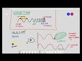
how are we able to see something if this is our eye you are only able to see something
if light was to hit the specimen so that's light over that light is able to hit the specimen and
the specimen must be able to reflect the light into your eyes then your photoreceptors in your eye detect the
light it sends an impulse to your brain and your brain goes
i'm able to see the specimen basically that's what's supposed to happen but let's talk about
what if the light was um let's say the object is like a transparent object if it's a transparent
object the light will just basically pass through and in that kind of situation the light
will not be reflected and we will not be able to see anything at all
that's what's supposed to happen so the first fundamental thing that you have to understand
is if you want to be able to see something if you want to be able to see something
the specimen interrupts the light and reflects the light
into our eyes that's the first thing we must understand so only when the light hits the specimen
and the light is reflected into our eyes we are able to see it fine now what's the big deal where does
magnification come in magnification comes in when you kind of put a lens
over here now when you put a lens over here what actually happens is it will go through the lens and in this
situation over here when it enters our eyes our eyes will comprehend the specimen to
be larger than it actually is this over here is the process of
magnification so this is the actual size of the specimen over here and this is the image size
pretty simple fine now and if we were to look at the actual size
over here if we were to make it bigger and this is the image size over here
this is just the act of magnifying it okay magnification equals you multiply the actual size and you'll
actually get the image size over here so that's how they get the formula
the formula equals to actual times magnification equals to image or you can also just rearrange it
where you can put magnification equals to image over actual that's how the formula comes to be so
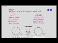
let's try a basic example basic example is as such if you have an actual size of a cell
which is about for example 10 micrometer and based on the
magnification of the cell we are certain the diameter of the cell was when we
looked at it under the microscope it was 10 millimeters so in this case they asked
you to calculate for example in the exam what is the magnification basically magnification okay i'm just writing it
like that so magnification equals to image over actual
and magnification equals to 10 millimeters which is the one that you are looking at
under the microscope okay what you're looking at what you see and 10 micrometer
is the actual size now a lot of students not a lot of students okay that's that's a bit of an unfair
statement to make but a few students have a tendency to just divide 10 millimeters to 10 micrometers
and they'll get the answer one and that is so wrong okay in maths the units for the numerator and the
denominator they have to be similar so you'll have to convert millimeter to micrometer and
to convert millimeter to micrometer you can just basically multiply it by a thousand
right and in this case it will be 10 000 micrometers divided by 10 micrometers and the answer in this case will be a
thousand that is the magnification right here okay
it's quite straightforward and of course the basic units that you'll have to know the units
in magnification uh rarely do we use centimeters but on the off chance it might come out
we have millimeters we have micrometer i'm not best at writing micrometer over there
okay and we also have a new one in igcse you only had to learn these three scales but you also have to introduce a
new one nanometer because we're going to be talking about organelles and organelles some
organelles fall under the scale of nanometers okay conversion of centimeters to millimeters
is multiplied by 10 millimeters to micrometers multiplied by a thousand
some books will put it as 10 to the power of three this to this micrometer to nanometer again multiply
10 to the power power of three or a thousand and nanometer to micrometer
it's the opposite you just divide it then divide the thousand divide a thousand over here that's a
comma by the way that's not a decimal place it has a separator all right and this one is divided by 10.
okay so these are the units that you'll have to know and you'll have to be able to
convert the units effortlessly in the exam all right they give you like for example three
millimeters okay three millimeters and they ask you to convert it into let's say micrometer so micrometer in
this case will be three millimeters times a thousand and therefore
it is equivalent to 3 000 micrometers some students do ask do i have to put the separator in the exam no you don't
have to it's just a habit um if you just basically want to adjust 3000 micrometer
go ahead no problems with that and if they do ask you to convert it into nanometer
then what you'll have to do is you'll have to do three multiplied by a thousand
multiply by another thousand and in this case it will be 3 million nanometers so
the conversion is something that we must be well versed with that is just basically what
magnification is all about magnification is just taking the actual specimen this is the
actual specimen and we magnify it using the lenses in the microscope so that when we
view it under the microscope what we are looking at is an enlarged image of the specimen
that is just what magnification is all about pretty simple and pretty straight
forward and the most important thing to also take away from this is if you want to be able to see
something if you want to be able to see something it must have the capability of interrupting the light
and reflecting the light this will be important for later and once it interrupts and reflects the
light it will then enter our eyes and therefore we are able to
comprehend the image that's how we look at it that's how we look at things on a daily
basis so for the next video what we're going to be looking at is
we're going to be seeing the wavelengths of a light microscope
The primary function of a microscope is to magnify the image size of a specimen, allowing us to see tiny structures that are otherwise invisible to the naked eye. This magnification enables detailed observation of cell components and their organization.
Magnification in microscopy occurs when a lens enlarges the image of a specimen. The formula for calculating magnification is Image size divided by Actual size, which helps determine how much larger the specimen appears under the microscope.
Key unit conversions for microscopy include: 1 centimeter = 10 millimeters, 1 millimeter = 1,000 micrometers, and 1 micrometer = 1,000 nanometers. Understanding these conversions is crucial for accurate magnification calculations.
Light interaction is essential because it allows the specimen to reflect light into our eyes. Transparent objects do not reflect light, making them invisible; thus, the specimen must interrupt and reflect light for us to perceive it.
After understanding microscopy and magnification, you can explore the wavelengths of light used in microscopes in the next video. Additionally, consider watching related content on cell structure and different types of microscopes to deepen your knowledge.
To calculate the magnification of a specimen, use the formula: Magnification = Image size / Actual size. For example, if a cell measures 10 micrometers and appears as 10 millimeters under the microscope, convert 10 millimeters to micrometers (10,000 micrometers) and then calculate: 10,000 micrometers / 10 micrometers = 1,000×.
For more information, check out videos like 'Understanding Cell Structure: The Amazing World Inside a Cell' for insights into cell components, or 'Understanding Microorganisms: Types of Microscopes and Their Applications' for a deeper dive into microscopy techniques.
Heads up!
This summary and transcript were automatically generated using AI with the Free YouTube Transcript Summary Tool by LunaNotes.
Generate a summary for freeRelated Summaries
Understanding Microorganisms: Types of Microscopes and Their Applications
Explore the fascinating world of microorganisms and the various microscopes used to observe them in detail.
Understanding Light Microscopy: Wavelengths and Visualization Limits
This summary explains how light microscopy uses different wavelengths of light to visualize specimens, highlighting why certain cellular structures like chloroplasts are visible while smaller ones like ribosomes are not. It covers key concepts such as light frequency, wavelength, and their biological significance in microscopy.
Understanding Light Microscope Resolution: Key Concepts Explained
This summary clarifies the complex concept of resolution in light microscopy, explaining how wavelength affects image detail and the ability to distinguish between close points. Learn why violet light offers better resolution than red light and the limitations of light microscopes in viewing extremely small specimens.

Understanding Cell Structure: The Amazing World Inside a Cell
Discover the wonders of cell structure, organelles, and their functions in this comprehensive guide.

Understanding the Structure and Function of the Cell: A Comprehensive Overview
Explore the intricate details of cell structure and function in our comprehensive guide, including organelles and their roles.
Most Viewed Summaries

A Comprehensive Guide to Using Stable Diffusion Forge UI
Explore the Stable Diffusion Forge UI, customizable settings, models, and more to enhance your image generation experience.

Kolonyalismo at Imperyalismo: Ang Kasaysayan ng Pagsakop sa Pilipinas
Tuklasin ang kasaysayan ng kolonyalismo at imperyalismo sa Pilipinas sa pamamagitan ni Ferdinand Magellan.

Mastering Inpainting with Stable Diffusion: Fix Mistakes and Enhance Your Images
Learn to fix mistakes and enhance images with Stable Diffusion's inpainting features effectively.

Pamamaraan at Patakarang Kolonyal ng mga Espanyol sa Pilipinas
Tuklasin ang mga pamamaraan at patakaran ng mga Espanyol sa Pilipinas, at ang epekto nito sa mga Pilipino.

How to Install and Configure Forge: A New Stable Diffusion Web UI
Learn to install and configure the new Forge web UI for Stable Diffusion, with tips on models and settings.

If you found this summary useful, consider buying us a coffee. It would help us a lot!