Understanding Light Microscopy: Wavelengths and Visualization Limits
Light microscopy uses visible light to visualize specimens. Although it involves some physics concepts, understanding light's properties is essential for biology students to grasp how microscopes work.
Key Concepts of Light in Microscopy
- Light as an Electromagnetic Wave: Light consists of waves with varying frequencies and wavelengths.
- Frequency: The number of wave peaks passing a point per unit time. High frequency means peaks are close together; low frequency means they are spaced further apart.
- Wavelength (λ): The distance between two consecutive peaks or valleys in a wave, measured in nanometers (nm).
Frequency and Wavelength of Visible Light
- Red Light: Low frequency, longer wavelength (~700 nm).
- Green Light: Medium frequency and wavelength.
- Violet Light: High frequency, shorter wavelength (~400 nm).
Why Wavelength Matters in Microscopy
- The ability to see a specimen depends on whether the light wavelength can interact with the specimen's size.
- Light must hit and reflect off the specimen to be visible under a microscope.
Visualization of Cellular Structures
- Chloroplasts: Large organelles (~2000 nm) that can interrupt and reflect light, making them visible under a light microscope. For more on the role of chloroplasts in biology, see Introduction to Microbiology: Understanding Microbes' Role in Life.
- Ribosomes: Much smaller (~23 nm), too small to interrupt light waves, so they cannot be seen with a light microscope. To learn more about cellular structures, check out Understanding Cell Structure: Basics of Microscopy and Magnification.
Practical Implications
- Light microscopes are limited by the wavelength of light used; structures smaller than the wavelength cannot be resolved. For a deeper understanding of the physics behind light, refer to Understanding Light: From Geometrical Optics to Quantum Mechanics.
- Understanding the relationship between wavelength and specimen size helps explain why some cellular components are visible and others are not.
Summary
- Light microscopy relies on light's wavelength and frequency to visualize specimens.
- Red light has a longer wavelength and lower frequency; violet light has a shorter wavelength and higher frequency.
- Larger organelles like chloroplasts can be seen because they reflect light, while smaller structures like ribosomes cannot.
- This knowledge is crucial for biology students, especially in exams like Cambridge A-levels, to understand microscopy limitations and applications.
light microscopy obviously like microscope the i mean it's in the name basically it uses light
okay it uses light to visualize a specimen now students hate this part a lot of
students especially hate this but they're like you'll be like oh my god i'm studying
biology why do i have to look at physics what does physics have to do with this because
we're gonna have to look at wavelength of light just a little bit so if you're not a physics student you
know uh you might have to just uh spend about a good five minutes for this
now light is an electromagnetic wave you don't have to memorize that for the exam but what you'll have to know is
different types of light have different frequencies light itself basically i'm not the best
at following this i'll find my best you have like different frequencies of light
so and i always love to ask my students which one is considered high frequency and which one's considered low frequency
uh if you are not so sure or if you know it let's just take some time let's just consider the
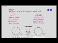
question now low frequency of light is this one where the peaks and the
valleys do not occur very frequently basically so it takes
some time for it to occur like this much of length there is only one peak and there's only
one valley we call that low frequency but high frequency is the peaks and the valley appear much
quicker we can just look at it that way you don't have to memorize what's the definition of low frequency and high
frequency but what you'll have to know is you'll have to kind of identify it if you are looking at a diagram which is
considered low frequency and which is considered high frequency now high frequency light i guess that's
purple yeah violet okay two certain extent my colors i'm not so good with my colors
this is high frequency it is extremely important to know that red light red light is considered a
low frequency light green falls somewhere in the middle over here
and violet is considered high frequency it should be just basically next to violet you must also know
the wavelength now when we're talking about the wavelength of light the wavelength of light is just
basically the distance between this peak to this peak
of this valley to this valley that's the wavelength of light it is usually symbolized as
lambda now so if they ask you the frequency of red light and the frequency of violet light
what do you think look at look at the distance between these two peaks over here and look at
the distance between these two peaks over here i'm just going to highlight it so that you can see it a bit more
clearly i'm just going to highlight these areas so who will have a higher wavelength
what do you think if you say that that light will have a higher wavelength you are correct
red light has a wavelength of about 700 nanometers and remember in the previous video
we did in introduce the concept of nanometers okay so that's a new one by the way and
violet light will have a frequency of about give or take
400 nanometers now why does this matter a lot of students are like why do i care about this why do i have
to give a damn about you know the length of what the wavelength of life how does this
matter in biology kind of does so in cambridge it is extremely important to know
for your a levels that red light has a frequency of 700 a wavelength of 700 nanometers it's a low frequency
wavelength and violet light has a 400 nanometer wavelength and its high frequency
all right now why is this a big deal or why do i care i love to ask my students these questions why
do i have to care about this okay how does this apply into microscopy or to visualize
a specimen now if you remember just a bit of revision from the
earlier video i mean the previous video the previous video i stated that if you want to visualize
an object okay or a specimen the light must first be able to hit the specimen
and reflect the specimen into i mean reflect the light into our eyes only then we will be
able to see it this is what i mentioned in the uh earlier video
now so let's apply this you see there are certain structures in a cell that are too small to be seen
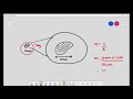
using light what do i mean by that let's consider the chloroplast now the chloroplast
itself is a very large organelle the size may vary but let's just say that the size of the organelle is
2 000 nanometers all right and let's also put another organelle over here
and that organelle i'm just gonna overhead the bottom is a ribosome okay and the ribosome over here is
actually only it should be smaller than that by the way so i hope maybe i should make it
into like a are you able to see this like like a green dot over there i hope you're able
to see that green dot and the ribosome is about give or take 23 nanometers let's just put 23
nanometers now imagine if i were to shoot light i don't care what color light i
shoot on it let's just say i just gave it red light now red light has a frequency of 700
nanometers so basically what will happen is it will just basically move along okay
and as the red light moves along does it hit the chloroplast
it hits the chloroplast and because it hits the chloroplast the chloroplast interrupts
light and reflects it so in this case if it's reflected will we be able to see it
the answer is yes you can use a light microscope and you can actually visualize the
chloroplast in a cell so in in other words you can actually view a chloroplast using a light microscope
now let's shift the focus to the ribosome which is about 23 nanometers
i'm going to ask you a question right now will we be able to see the ribosome using red light
or violet light for that matter will we be able to see it the answer is probably no because
what actually happens what do you notice what is happening over here exactly the red light just passes
through the ribosome the ribosome does not have the capability of interrupting the light so how now
can so can we see ribosomes using a light microscope no we can't we will not be able to visualize a
ribosome because they are too small
Light microscopy is a technique that uses visible light to visualize specimens. It relies on the properties of light, such as wavelength and frequency, to illuminate and magnify objects, allowing us to see cellular structures that are otherwise invisible to the naked eye.
The wavelength of light is crucial in microscopy because it determines the ability to resolve details in a specimen. Structures smaller than the wavelength of light cannot be seen, as they do not effectively interact with the light, which is why larger organelles like chloroplasts are visible while smaller structures like ribosomes are not.
Different colors of light correspond to different wavelengths and frequencies. For example, red light has a longer wavelength (~700 nm) and lower frequency, while violet light has a shorter wavelength (~400 nm) and higher frequency. This variation affects how well certain specimens can be visualized under a microscope, with shorter wavelengths generally providing better resolution.
Examples of cellular structures visible under a light microscope include chloroplasts, which are large enough (~2000 nm) to reflect light and be seen. In contrast, smaller structures like ribosomes (~23 nm) cannot be visualized because they are below the resolution limit of light microscopy.
The primary limitation of light microscopy is its inability to resolve structures smaller than the wavelength of light used. This means that while larger organelles can be seen, smaller components like ribosomes cannot be visualized, which can limit the understanding of cellular functions.
Understanding light microscopy is essential for biology students as it helps them grasp the limitations and applications of this technique in studying cells. This knowledge is particularly important for exams, such as Cambridge A-levels, where a solid understanding of microscopy principles is tested.
For a deeper understanding of the physics behind light and its applications in microscopy, you can refer to resources like 'Understanding Light: From Geometrical Optics to Quantum Mechanics.' This will provide insights into how light behaves and its implications for microscopy.
Heads up!
This summary and transcript were automatically generated using AI with the Free YouTube Transcript Summary Tool by LunaNotes.
Generate a summary for freeRelated Summaries
Understanding Light Microscope Resolution: Key Concepts Explained
This summary clarifies the complex concept of resolution in light microscopy, explaining how wavelength affects image detail and the ability to distinguish between close points. Learn why violet light offers better resolution than red light and the limitations of light microscopes in viewing extremely small specimens.
Understanding Cell Structure: Basics of Microscopy and Magnification
This video explains the fundamentals of cell microscopy, focusing on how microscopes magnify specimens to reveal cell structures. Learn about light microscopes, the concept of magnification, unit conversions, and how light interaction enables us to see microscopic objects.
Understanding Microorganisms: Types of Microscopes and Their Applications
Explore the fascinating world of microorganisms and the various microscopes used to observe them in detail.
Understanding Light: From Geometrical Optics to Quantum Mechanics
Explore the evolution of light theory from Maxwell's equations to the concept of photons in quantum mechanics.

Introduction to Microbiology: Understanding Microbes and Their Importance
Explore the fascinating world of microbiology, learn about various microbes and their significance to life on Earth.
Most Viewed Summaries

A Comprehensive Guide to Using Stable Diffusion Forge UI
Explore the Stable Diffusion Forge UI, customizable settings, models, and more to enhance your image generation experience.

Kolonyalismo at Imperyalismo: Ang Kasaysayan ng Pagsakop sa Pilipinas
Tuklasin ang kasaysayan ng kolonyalismo at imperyalismo sa Pilipinas sa pamamagitan ni Ferdinand Magellan.

Mastering Inpainting with Stable Diffusion: Fix Mistakes and Enhance Your Images
Learn to fix mistakes and enhance images with Stable Diffusion's inpainting features effectively.

Pamamaraan at Patakarang Kolonyal ng mga Espanyol sa Pilipinas
Tuklasin ang mga pamamaraan at patakaran ng mga Espanyol sa Pilipinas, at ang epekto nito sa mga Pilipino.

How to Install and Configure Forge: A New Stable Diffusion Web UI
Learn to install and configure the new Forge web UI for Stable Diffusion, with tips on models and settings.

If you found this summary useful, consider buying us a coffee. It would help us a lot!