Introduction
Microorganisms, despite their minuscule size, play pivotal roles in ecosystems and human health. Yet, their smallness means they can’t be observed with the naked eye. To study these tiny life forms, scientists rely on important tools known as microscopes. The word 'microscope' is derived from Latin, where 'micro' means small, and 'scope' indicates to look at or view. In this article, we will explore the various types of microscopes, their functionalities, and how they enable us to observe microorganisms in incredible detail.
Types of Microscopes
Understanding microorganisms requires various techniques of microscopic examination, which can be broadly divided into two categories: light microscopy and electron microscopy. Each category encompasses several types of instruments, each designed for specific observations.
Light Microscopy
Light microscopy utilizes visible light to magnify specimens. It is the most commonly used method in laboratories for observing microorganisms. Here are the main subtypes:
Compound Light Microscopes
These microscopes employ two or more lenses, allowing for significant magnification.
- Objective Lenses: Located near the specimen, they further magnify the image.
- Ocular Lenses: The eyepiece, where the image is viewed, adds additional magnification.
For example, if an objective lens magnifies 4x and the ocular lens magnifies 10x, the total magnification is 40x (4 × 10).
Dark Field Microscopes
This variation illuminates the specimen with light that does not enter the objective directly. Instead, the light is reflected off the specimen, appearing brighter against a dark background. This is particularly useful for observing live microorganisms that cannot be stained.
Phase Contrast Microscopes
Phase contrast microscopes are used to enhance the contrast of transparent specimens. They utilize ring-shaped diaphragms to create dark backgrounds, enabling detailed examination of living cells without staining.
Differential Interference Contrast Microscopes
Similar to phase contrast, this type uses two beams of light split by prisms. This results in high-contrast images that appear three-dimensional, enabling the viewer to observe internal structures of living specimens.
Fluorescence Microscopes
These microscopes leverage ultraviolet light to excite fluorescent dyes attached to specimens. This method is effective for rapid identification of bacteria and other microorganisms, providing vibrant images against dark backgrounds.
Confocal Microscopes
Confocal microscopy uses lasers to illuminate specimens, capturing images from various depth planes. This results in high-resolution, three-dimensional images of structures within cells, useful in biomedical research.
Electron Microscopy
For viewing structures smaller than 0.2 micrometers, like viruses, electron microscopes are employed. They use electron beams for imaging, providing much greater resolution than light microscopes.
Transmission Electron Microscopes (TEM)
TEM focuses a beam of electrons that passes through thin sections of a specimen, producing two-dimensional images with exceptional resolution. This type is vital for studying organelles in cells.
Scanning Electron Microscopes (SEM)
In contrast to TEM, SEM scans the surface of a specimen with electrons, yielding three-dimensional images. This is ideal for studying the surface features of microorganisms and cells.
Scanning Probe Microscopes
These innovative microscopes use probes to scan specimens, providing a view of molecular structures without damaging the sample. The two primary types include:
- Scanning Tunneling Microscopes (STM): With exceptionally high resolution, STMs can visualize structures at the atomic level.
- Atomic Force Microscopes (AFM): AFMs, using a metal and diamond probe, record movements as they pass over surfaces, producing three-dimensional images.
Measuring Microorganisms
When discussing microorganisms, we frequently reference their size via specific units:
- Micrometers (µm): One micrometer is one millionth of a meter, typically used to measure the length of bacteria.
- Nanometers (nm): One nanometer equals one billionth of a meter, often utilized for viruses and cellular structures.
Understanding these measurements is crucial because microorganisms exist at a scale that necessitates the use of such small units—highlighting the importance of precise methods like microscopy in microbiology.
The Importance of Resolution in Microscopy
Resolution, when it comes to microscopy, refers to the ability to distinguish two points as separate. This aspect is crucial for effectively viewing microorganisms. Various microscopy techniques improve resolution in different contexts:
- High-resolution examples: Those derived from electron microscopy allow us to observe fine details within cells, such as organelles and fibers.
- Challenges of Lower Resolution: Using lower resolution methods may render cellular structures indistinct, leading to blurry images that hinder our understanding.
Conclusion
In summary, microscopes are essential tools in microbiology that open up the hidden world of microorganisms. Different types of microscopes, whether they use light or electron beams, serve varied purposes and allow for the exploration of life forms at scales previously unimaginable. Through advancements in microscopy, researchers can not only visualize these entities but also advance our understanding of their roles within ecosystems and diseases. By integrating knowledge of measurement techniques and resolution capabilities, scientists continue to peel back the layers of intricacy that define the microbial universe.
microorganisms are way too small to be seen with the unaided eye instead we'll need to use a microscope to view them
microscope is a term that is derived from latin we can break it down into micro which means small
in scope to look at we're going to take a look at the different types of microscopes that
exist those that are ancient using those single lenses to view and magnify
organisms to those that can use electron beams to magnify them instead when it comes to
units of measurement we use the metric system in the sciences especially in the medical field and of
course the major advantage here of the metric system is that it relates to the other
unit and factors by 10 which means that when we move from one unit to the next we're usually
multiplying or dividing by a factor of 10 either by 10 100
a thousand so on so forth so if we take a look at meter which is the basic unit for length
and we try to use that in the context of measuring the size of these different organisms we
will be able to measure them in the micrometer range and some even in the nanometer
so microorganisms are measured in very small units micrometers and nanometers so
how does that compare to meters well a micrometer one micrometer equals ten to the negative six meters the
prefix micro indicates that this unit meter should be divided by 1 million
or 10 to the power 6. if we take a look at nanometers then we need to divide the meter unit by
a billion or 10 to the power 9. so in this case we're using very small units and prefixes to describe the size
of microorganisms one micrometer for bacteria
and in the nanometers for viruses we use microscopes to aid us in viewing those microorganisms
and we have different types so in general we either have simple microscopes that
use one lens or compound microscopes that use more than one
lens the simplest microscope used by van leuven hoax had only one lens it was kind of similar to a magnifying glass
but it had much better magnification then after that we had robert hook who built compound microscopes those
that have multiple lenses and then several after kept building and making those microscopes better
of course those early microscopes were of poor quality and were not used to see bacteria
it wasn't until the 1830s until better microscopes were developed by the likes of lister
and much improvements were made to these microscopes to allow for the viewing
of microscopic organisms like bacteria but our focus here are compound microscopes
that use multiple lenses to view specimen and under compound microscopes we have
several different types we have light microscopes two photon microscopes scanning acoustic
of course we then have the electron microscopes and the scan probe microscopy compound microscopes are also
known as light microscopes and we're going to focus on several examples
of this type of microscope first and then discuss the rest
so starting with light microscopy light microscopy we have several different
subtypes that use light visible light to observe specimen any kind of this type of microscope is
classified under light microscope so compound like microscopes dark field face contrast
differential interference fluorescence and confocal microscopy are all examples of light microscopes
the term compound microscopes again refer to the fact that they used two lenses
to view the organism and it's probably the one that you're most familiar with or have seen in the
lab the compound light microscopes lenses are housed in the objective area
and in the ocular piece and the setup inside is magnify once using the objective lenses
magnify another time using the ocular lens and that magnification is multiplied
so if you have here an objective that magnifies your image by four times and then the
ocular piece magnifies the image by ten times then the total magnification
needs to be multiplied four times ten the total magnification of your image would be 4dx
and that's what compound light microscopes are they use more than one lens
to magnify the image the specimen is placed on a stage that can be lowered or raised depending
on what objective we're using and the light source is
on the bottom so the light source will direct the visible light through a condenser and a
diaphragm that focuses the light into the specimen in the slide through the lens
of the objective specific lens that you're using and then through this whole area
and then the last set of lenses which are the oculars and then allows you see to see the image
through the eyepiece in a compound microscope the image that forms is magnified like i said twice so
the total magnification we'll need to consider the objective lens
times the ocular lens one term that you hear frequently when talking about microscopes is resolution
what does this mean resolution refers to the ability of the lens to distinguish two points
and you can see here with this image that as we go through the different images
we are able to see the letter more clearly this magnification
and this image has great resolution what does this mean so if you think about
let's say a highway or you're driving on a road that has two-way traffic and you're driving at night and you're
able to see the cars that are coming towards you in your direction
and then passing you as you drive on this two-way street when the car is still far
away you might see the headlights as just one big
blob of light coming towards you but as you get closer and closer to the car you can see that there are two distinct
headlights as you come closer to it and that's kind of the same idea if a microscope has better resolution
you're going to be able to differentiate different spots in your specimen as distinct
areas if it has poor resolution then you're just not going to be able to distinguish those two items as separate
items instead they're just going to look like one blurry image and
we're going to see here with different types of microscopes different magnifications and different
types are going to have different types of resolution another thing that i want to point out when it
comes to compound light microscopes is the refractive index the refractive index measures
the light bending ability of a medium you've probably seen this experiment where you have water in a clear glass
and then you put a pencil in that glass and you look at the interface where
water and air meet you try to focus on the image of the pencil you will see at that point that the
image is a little distorted as soon as you look at the image in the water it's going to look like the
pencil is towards the side not continuous and that is due to the light
moving from one medium to the other which is air into liquid or liquid into air
and in this case light bends as it moves through this these different media
and the same is going to happen when the light is moving from the light source through the air through the glass
through the air again and into the objective lens there's going to be
some bending of the light and when light bends and you're not able to capture it that's going to make
the specimen blurry when does this become a problem this becomes a problem with magnifications
that are pretty high 100x for example is the highest magnification
on a bright field compound light microscope and at that point the refractive index
is pretty big you can take a look here how much light bends when we're using the 100x
lens so the light will not be able to pass through here as efficiently and we're going to
see a blurry image so how do we fix this we add a drop of oil drop this drop of
oil is going to reduce the bending ability of the medium and in this case the light
will be able to continue to pass through the objective lens
and make the image a little bit clearer take a look here in the air when light is passing through this solid
to this air medium the refraction is great light will not be able to pass through
however adding immersion oil will allow the light to bend less and continue forward through
the objective lens allowing us to see a better image so this is the basic setup for a
compound light microscope we're going to see several different examples that have
slight variations so what i've been talking about so far is the compound light microscope that can
use bright field elimination the path of light in bright field microscopy
is just using regular visible light from the bottom so going back here this is the light source through the
specimen and then the light is collected through the objective lens
now as the light passes through the dark objects will be visible against the bright background
light will be reflected of the specimen and the light that is reflected does not enter
the objective lens so all in all characteristics of this type of microscope
is that it uses visible light as a source of elimination and the best resolution we can get is
around 0.2 micrometers and what you're going to see is a specimen
that appears against a bright background so the specimen will look dark and the surrounding will look bright
some advantages of this microscope is that it's pretty inexpensive it's easy to use
we use it to observe stained organisms in the lab however it does not allow us to view
very small specimens like viruses the next major type of microscope light
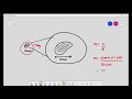
microscope is the dark field microscope this one uses a special condenser with an opaque disc
that illuminates all light in the center of the beam what does this mean
it means that light is reflected by the specimen and will enter the objective lens what we will see
is in this case a specimen that looks light and the background will look dark
so if you're using this microscope your problem this phase of the microscope is because
the microorganism that you're viewing might be
live or if you were to use a regular microscope it might look invisible because of how light
it is you cannot stain a microorganism maybe or it could be a ruined if you do stain
it so what this microscope does is it uses an
opaque disc and when i say opaque it means black dark and so we insert that disc
and we allow the light to pass the disc is going to block that light and it's not going to enter the objective
lens directly the only light that is going to be reflected is going to be reflected
from the specimen and that's going to enter the objective lens
because there's no direct background light the specimen will look light
against that black background the next example here is to show you what the opaque disc does
and how it blocks the light from entering the objective directly and instead all the light that is entering
is from the specimen itself and you can see it here this is the light source this is the
opaque disk and this is a specimen so really this is mostly dark the
background is going to be dark the light that is reflected off of the specimen is collected
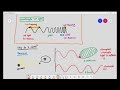
through the objective next one is face contrast face contrast uses a special condenser
and it has a ring-shaped diaphragm the diaphragm's function is to allow direct light to
pass through the condenser focusing the light on the specimen and then a diffraction plate
is also used in the objective lens we have two types of light rays we have direct light rays
and reflected light rays they're both brought together to produce an image why would we use this type of microscope
this allows us to see detailed examination of the internal structure of
living specimen and you can see that with the image right here so
the main function of this microscope is to bring together two sets of light rays direct light rays and reflected or
diffracted and the image that is collected will allow us to examine
detailed inner structures so if you look at the phase contrast type of microscope setup
you can see that there are different phases between the light that is passing
through the object and the background and we superimpose the light rays producing a contrast so the light source
is here this is the diaphragm ring that i was mentioning
and in here we have different types of light that are passing
through the specimen and then the specimen will reflect certain type of light the light that is
traveling directly from the condenser lens and the light that is traveling from the
specimen are going to be out of phase and when that happens
that image that is collected will allow for viewing of internal structures
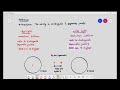
the fourth major type of microscope under the light microscope category is differential interference contrast
this is similar to phase contrast however it uses differences in refractive indices
to produce images so it uses two beams of light that are separated by prism so the
specimen will appear like it's colored because of this prism effect and what's cool about this microscope is
it allows you to see the three-dimensional image of the specimen that you're
viewing so the major idea here is that it uses two beams of light and there's a prism
in there it's going to split those light beams giving more contrast and color to the
specimen and allowing you to view this in 3d the last type of microscopy under light
category is a fluorescence microscopy and this type of a microscope uses an ultraviolet source of illumination
that will cause fluorescent compounds in the specimen to emit light so those fluorescent substances are
usually fluorescent dyes that can stain the cell especially if the cells do not naturally
fluoresce and using this microscope the dyes will give off color
when viewed under this microscope and the uses here are for rapid detection and identification of
microorganisms in clinical specimen or tissue for example those
fluorochromes those dyes they have special attraction to different organisms and
as an example one of them glows yellow when we expose it to uv light and this type of
fluorochrome is absorbed by a bacteria we call mycobacterium tuberculosis the bacteria that causes tuberculosis
and when we apply this dye to the sample especially if we suspect
that the bacteria is there um it's going to allow us to see a bright yellow
organism against a dark background another one is the organism bacillus and thrices
the causative agent for anthrax it appears apple green when we stain it with another
fluorochrome so far the different types of microscopes that we mentioned are under
the light microscopy category we're going to move to the next category which discuss confocal microscopy
confocal microscopy is similar to the last one which we just talked about the fluorescence
microscope and this one specifically also uses fluorochrome dyes
however the microscope employs the use of a single photon to illuminate a plane of specimen at a time what that
means is it's going to kind of section it and then it's going to illuminate every
section or each plane of the specimen and it will allow us to see the three-dimensional shape
especially that it also emits those fluorescent colors what would we use this for
this would be used to obtain 3d dimensional images of cells especially for biomedical
applications it allows us to view biofilms as you're seeing with this image
and we can use them to directly stain different types of bacteria that cause infection
like niceria gonorrhea the next type of microscope is a two photon microscope currently
using those microscopes is limited to very advanced research and clinical labs because of
how much they cost and a single two photon microscope usually costs between three hundred
thousand to five hundred thousand dollars because it uses those lasers to
excite the dyes that we use on the specimen for this specific type of microscope
similar to confocal microscopy the specimen is stained with those fluorochromes those fluorescent dyes
however the two-photon microscope uses two types of photons uses a long wavelength light
and the two photons that are then used will excite the dyes to emit light
this allows us to view the depth of living cells that are around
one millimeter so it actually allows us to see cells activity in
real time for example cells of our immune system have been observed using this microscope
as they respond to an antigen the next category are the super resolution light microscopes
and those microscopes use two laser lights to illuminate one nanometer at a time it also uses
fluorescent molecules to glow the specimen and the second wavelength is going to cancel all the fluorescence
except for that one nanometer that it's supposed to illuminate
the major purpose of this type of microscopy after putting all the images together
is to observe the location of the different molecules inside the cell next up we have the
scanning acoustic microscopes also called sam this basically consists of interpreting the action of sound
waves that are sent through the specimen so instead of actually sending light or photons
sound waves are sent through they have specific frequency they travel through the specimen
and some of it is reflected and when it hits the interface within the material then
all of that is collected and translated into an image using a computer the this type of microscope is used to
study living cells that are attached to a surface especially cancer cells or artery
plaques and even bacterial biofilm another category of microscopes are electron
microscopes these types of microscopes use electron beams
instead of visible light to view the specimen especially objects that are smaller than
0.2 micrometers like viruses and internal structures of cells all of that can be viewed using this
microscope and these free electrons travel in waves are collected by the use of a magnet
so if you take a look at the internal setup of an electron microscope and compare it to a light microscope
you'll notice that these electron microscopes use magnets to focus the electron beams
very similar to how light microscopes use lenses to focus light it's important to
note that there are two main types of electron microscopes transmission electron microscopes
and scanning there are differences between the two in the electron microscope specifically
the transmission one the beam of electrons is focused using an electron gun and it will
pass through the specimen that is prepared using a very ultra thin section
of that specimen the beam is then focused on a small area of the specimen through a condenser lens
directing the electron beams in a straight line to illuminate the specimen
so you can see the electron gun here the condenser we have a second condenser we have the specimen and then the
electrons are focused by an electromagnetic projector lens onto
a screen final image that we call a micrograph will appear as many light and dark areas
depending on how many electrons are absorbed by the different areas of the specimen
the transmission electron microscope has very high resolution it allows us to see the
organism or the specimen in a 2d manner so if you're looking for that information you'll find it on this slide
and in order to use the transmission electron microscope and view the specimen the specimen has to be stained
with metal salts and the magnification is between 10 000 to 10 million
for the scanning electron microscope it uses this beam of electrons to scan the surface of the entire specimen
the scanning electron microscope will use a beam of electrons that is produced from an electron gun uh
it will use this electromagnetic lens to direct the electron through the surface of the
specimen and the the image that is produced will be viewed on a screen
now what's uh special about the scanning electron microscope is it allows you to see the 3d
structure of the organism because it's only going to direct those electrons to the surface and it
allows the magnification to be around 1000 to 500 000. so the the main purpose of the scanning
is to study the surface features of cells and viruses the transmission electron microscope
allows you to see the inside and will need to use very thin slices so you can compare the two
images that you can see so for example this is the image of a cell in a biofilm you can see
some internal structures because of how the transmission electron microscope works
however the scanning only scans the surface so you can see the 3d structure of staphylococcus aureus
in this image the last category of microscopes that i want to discuss are the scanning probe microscopes since
the early 1980s several of these microscopes have been developed
they use probes to examine the surface of a specimen using electric currents they don't
modify the organism or expose it to any damaging radiation
these microscopes can get very close to the molecular shapes inside the cell it can characterize our
chemical properties it can determine temperature in different areas of the cell and we
have two major kinds we have the the tunneling microscope and the atomic force
microscope the tunneling one specifically uses a tungsten probe that scans
the specimen and produces an image that reveals the bumps and the depressions of the atoms on the surface
and basically giving us great details about its surface the resolution here is very powerful
it's even greater than that of an electron microscope it can resolve features that are about 1
over 100 the size of an atom take a look here this is an image produced by the scanning tunneling
microscope of pure gold surface we're looking at the atoms of gold
and how they are arranged this image is of an atomic force microscope
the atomic force microscope uses a metal and diamond probe that is gently forced down onto the
specimen and it moves along the surface and the movements are recorded we can see the 3d
image that is produced this image here shows long stranded like molecules
of cellulose that is derived from from plant fibers all of these different types of microscopes that we
talked about can be used in microscopy we've discussed them in the order of increasing
resolution our most used microscopes are the light microscopes especially in science labs
and the rest are for more clinical purposes and are more expensive
Heads up!
This summary and transcript were automatically generated using AI with the Free YouTube Transcript Summary Tool by LunaNotes.
Generate a summary for freeRelated Summaries

Introduction to Microbiology: Understanding Microbes and Their Importance
Explore the fascinating world of microbiology, learn about various microbes and their significance to life on Earth.
Introduction to Microbiology: Understanding Microbes' Role in Life
Explore the world of microbes, their classifications, and their importance in sustaining life on Earth.
Understanding Cell Structure: Basics of Microscopy and Magnification
This video explains the fundamentals of cell microscopy, focusing on how microscopes magnify specimens to reveal cell structures. Learn about light microscopes, the concept of magnification, unit conversions, and how light interaction enables us to see microscopic objects.
Understanding Light Microscopy: Wavelengths and Visualization Limits
This summary explains how light microscopy uses different wavelengths of light to visualize specimens, highlighting why certain cellular structures like chloroplasts are visible while smaller ones like ribosomes are not. It covers key concepts such as light frequency, wavelength, and their biological significance in microscopy.
Understanding Light Microscope Resolution: Key Concepts Explained
This summary clarifies the complex concept of resolution in light microscopy, explaining how wavelength affects image detail and the ability to distinguish between close points. Learn why violet light offers better resolution than red light and the limitations of light microscopes in viewing extremely small specimens.
Most Viewed Summaries

A Comprehensive Guide to Using Stable Diffusion Forge UI
Explore the Stable Diffusion Forge UI, customizable settings, models, and more to enhance your image generation experience.

Kolonyalismo at Imperyalismo: Ang Kasaysayan ng Pagsakop sa Pilipinas
Tuklasin ang kasaysayan ng kolonyalismo at imperyalismo sa Pilipinas sa pamamagitan ni Ferdinand Magellan.

Mastering Inpainting with Stable Diffusion: Fix Mistakes and Enhance Your Images
Learn to fix mistakes and enhance images with Stable Diffusion's inpainting features effectively.

Pamamaraan at Patakarang Kolonyal ng mga Espanyol sa Pilipinas
Tuklasin ang mga pamamaraan at patakaran ng mga Espanyol sa Pilipinas, at ang epekto nito sa mga Pilipino.

How to Install and Configure Forge: A New Stable Diffusion Web UI
Learn to install and configure the new Forge web UI for Stable Diffusion, with tips on models and settings.

If you found this summary useful, consider buying us a coffee. It would help us a lot!