Understanding Light Microscope Resolution: Key Concepts Explained
Introduction to Magnification and Resolution in Light Microscopy
- Magnification enlarges the specimen size but does not improve image detail.
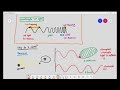
- Light wavelength ranges from 400 nm (violet) to 700 nm (red), which influences resolution.
What is Resolution?
- Resolution is the ability to distinguish or separate two close points in an image.
- It also refers to the sharpness or detail of the image.
- The smaller the resolution value (in nanometers), the better the microscope can distinguish details.
Calculating Resolution
- Resolution (R) = Wavelength of light / 2
- Example: Using violet light (400 nm), resolution = 400/2 = 200 nm.
- Using red light (700 nm), resolution = 700/2 = 350 nm.
Practical Examples of Resolution
-
Two points 500 nm apart:
- Both red and violet light microscopes can distinguish two separate points.
- Image appears detailed and clear.
-
Two points 300 nm apart:
- Violet light (200 nm resolution) can distinguish two points.
- Red light (350 nm resolution) cannot; image appears as a single blob.
-
Two points 100 nm apart:
- Neither red nor violet light microscopes can resolve the points.
- Image appears as one indistinct blob.
Why Resolution Matters
- Light microscopes cannot resolve objects smaller than their resolution limit (about 200 nm for violet light).
- Ribosomes, for example, are too small to be resolved by light microscopes.
- When points are closer than the resolution limit, images lose detail and sharpness.
Comparing Red and Violet Light
- Violet light has a shorter wavelength, thus better resolution and sharper images.
- Red light has a longer wavelength, resulting in lower resolution and less detailed images.
Limitations of Light Microscopes
- Cannot resolve objects smaller than approximately 200 nm.
- Cannot clearly distinguish points closer than the resolution limit.
- Scientists need more advanced microscopes (e.g., electron microscopes) to study very small structures.
Summary
- Magnification enlarges images but does not improve resolution.
- Resolution depends on the wavelength of light used.
- Violet light provides better resolution than red light.
- Light microscopes have inherent limits in resolving very small or closely spaced objects.
- Understanding resolution helps in choosing the right microscopy technique for detailed specimen analysis.
For a deeper understanding of how light microscopy works, you may find the following resources helpful:
- Understanding Light Microscopy: Wavelengths and Visualization Limits
- Understanding Cell Structure: Basics of Microscopy and Magnification
- Understanding Microorganisms: Types of Microscopes and Their Applications
- Understanding Light: From Geometrical Optics to Quantum Mechanics
- Understanding Spherical and Chromatic Aberration in Optical Lenses
now this is where it gets a little bit confusing
what we did earlier was we talked about magnification and we said that magnification is just
basically to enlarge the specimen size and we also said earlier
that light has a wavelength of about 400 nanometers to 700 nanometers just a bit of revision 400 nanometer is
violet 700 nanometer is red light always never lose sight of that you must
remember that now comes the most i would say for this chapter at least the most confusing
concept which is the concept of resolution a lot of students especially when i'm
teaching them they'll look at a textbook definition and they'll be like yeah i don't understand that what does
that even mean now resolution in the textbook basically says the ability
to distinguish or separate between two separate points and a lot of students just basically
go okay what does that even mean uh oh another definition that you can also include in the exam
is the sharpness or detail of an image so what so let's understand or let's
explore this concept a little bit a very important thing to understand about the
resolution of light microscopes is basically this the resolution of light microscope
or i'm just going to put resolution las equals to the wavelength of the electromagnetic
wave divided by two so if they ask you to calculate the resolution
okay the resolution using violet light it basically means 400 divided by 2
equals to 200 nanometers that's a very simple calculation but it does not answer the question what
does it even mean that it has a resolution of 200 nanometers
and for resolution the smaller the number the better i'll explain why
let's imagine a situation over here and the situation is as such you are viewing a specimen under a
microscope and the specimen is as such the actual specimen
looks very uh simple it is just a two simple dots and the two simple dots over here are
500 nanometers apart we're going to ask a very simple question over here
what do we see yes this is very important okay what do we see do we see as two
separate spots or do we see it as one spot if you're using a light microscope yes
you will still see it as two separate points now why do you see it as two separate points
the reason you see it as two separate points is because light
if you remember has a maximum resolution it's going to put max os of 200 nanometers
so anything 200 nanometers and above light is able to distinguish between the two
separate spots over here now if you don't understand this let's try another example
the actual specimen is now ha this is where it becomes interesting the actual specimen is now 100
nanometers above but what do we see using in our eyepiece when we are viewing it under the
eyepiece over here we are looking at it do we see it as two separate spots or do we see it as one spot
see here's where it becomes a little odd you will only be able to see it as a single blob over here now why is that
so why don't you see two separate spots is that detailed no it's not detailed
because this is the detailed image but this one over here what you see is not detailed at all
what gives why does this happen this happens because the two spots are less than
200 nanometers apart and because they are less than 200 nanometers apart what happens
light waves are unable to resolve the image therefore in this case it is less sharp or basically less
detailed that's what that means so we have a problem problem number one light microscope over
here it's unable to visualize extremely small specimens
an example of this extremely small specimen that i told you before was the ribosome because why couldn't it
resolve the ribosome because the ribosome was too small and remember the light wavelength
it just basically the ribosome is in this case the ribosome is unable to interrupt light
so we have the first problem over here number two it's unable to resolve
any specimen smaller than 200 nanometers now a lot of students think that if you're not able to resolve
anything smaller than 200 nanometers it means you're not able to see it uh it does not mean that you might still
be able to see something like for example let's say let's say in this case you
have two specimens over here you have two specimens over here under the microscope the only issue is the two
specimens are 180 nanometers apart but the problem is when you're using the light microscope
what we might see is we might just see it as a single blob over here so it is less detailed so it's
not sharp you're not able to study the specimen in detail that's the problem with light
microscopes they they work they have some advantages by the way i'm not i'm not like you know bashing light
microscopes there is a time and place to use light microscopes definitely
however they do have their own shortcomings so we're going to be talking about
the differences in resolution between red light and violet light because
sometimes students get a little bit confused when they're comparing the resolution between
two different colors now this is a bit of reference for you what is the meaning of resolution by the way
resolution is just basically the ability to distinguish between two separate points that's what we
discussed earlier and remember red light will have a resolution of 350 nanometers
how did i get 350 nanometers if you remember red light has a wavelength of 700
nanometers and it's divided by two that is the formula so what does it mean by
a resolution of 350 nanometers this means that red light has the ability to distinguish
two separate points which are 350 nanometers and higher violet light however has a resolution of
200 nanometers and if you remember violet light has a wavelength of 400 nanometers divided by 2 and you
will get a resolution of 200 nanometers basically it means that
violet light is able to distinguish separate points of 200 nanometers and higher
and i did ask uh in an earlier question which light has a better resolving power or which light
provides better resolution and i did say that violet light is better than red light
in producing a more detailed image under the microscope how is that so let's see
here we have two circles and the circles basically show us our eyepiece of the light
microscope this is our eyepiece of the light microscope and we will be viewing a particular
image based on a natural specimen now the actual specimen is let's for
example just say there are two bacteria over here the bacteria are those orange color
circles over there okay there you can see two bacteria and for example these two bacteria uh
i'm just gonna put a distance these two bacteria are 500 nanometers apart if
we view this bacteria under the light microscope using red light will we see
two bacteria or will we see a shapeless blob we will see two bacteria over here for
the red light and we will also see two bacteria for the violet light so this is
say to be a detailed image why does this mean that this is a detailed image because
in reality there were two bacteria which are 500 nanometers apart and when we viewed it under the
microscope we are able to see the two bacteria so the image is clear now why are we able to see two bacteria
using red light or using violet light the reason is because like i said earlier
if you remember red light has a this ability red light has a ability a red light has an ability
to distinguish between points which are 350 nanometers and higher so because the bacteria is
500 nanometers apart red light has the ability to distinguish them so is violet light by the way
what if we have two bacteria viewed under the microscope and the bacteria are actually 300 nanometers apart this
is where it becomes interesting if you were viewing this specimen under red light
you will not be able to see the two bacteria instead you will see a shapeless mass
you're unable to distinguish the two separate points over here so you will not be sure of what you're
exactly looking at are you looking at one bacterium or are you looking at two bacteria in
this case reason is because the actual distance of the bacteria is
300 nanometers and red light can only resolve things which are 350 nanometers and higher
anything below 350 nanometers it like becomes a little bit useless you're still able to see something but
you're not exactly sure what you're looking at that's why we say it's less detailed however with violet
light you're still able to see it as two separate points you are able to see hey there are two bacteria over there
because their distance is 300 nanometers apart and violet light can distinguish
anything 200 nanometers apart and higher so in this case red light gives you a less detailed image
violet light gives you a more detailed image even though both of them are being
viewed on the same magnification and let's try one last experiment over here now we have two bacteria
being viewed under the microscope and the two bacteria are about 100 nanometers apart
if we were to view this bacteria under red light we will see it as one shapeless blob
and if we were to view it under violet light we will also see it as one shapeless blob
we are not exactly sure what we are looking at why is that so because the actual
distance of the two bacteria are 100 nanometer apart red light has a resolution of 350
nanometers and higher violet light has a resolution of 200 nanometers and higher
both these lights are unable to resolve anything lower than 200 nanometers and therefore
they are unable to distinguish two separate points which are 100 nanometers apart
so in this situation because the distance of the bacteria is 100 nanometers apart using light
microscope will give you a less detailed image and scientists in this case will be
concerned because they won't they will be wondering am i actually looking at
one bacterium am i looking at two bacteria what exactly am i looking at light microscope has now reached its
limit and we need a better microscope we need a better solution
Magnification refers to the enlargement of a specimen's size, while resolution is the ability to distinguish two close points in an image. Higher magnification does not necessarily mean better detail; resolution is crucial for clarity and sharpness in the observed image.
Resolution can be calculated using the formula: Resolution (R) = Wavelength of light / 2. For example, using violet light with a wavelength of 400 nm, the resolution would be 200 nm, while using red light at 700 nm results in a resolution of 350 nm.
The wavelength of light affects resolution because shorter wavelengths can distinguish smaller details. Violet light, with a shorter wavelength, provides better resolution than red light, allowing for clearer images of closely spaced points.
Light microscopes cannot resolve objects smaller than their resolution limit, which is about 200 nm for violet light. This means that structures like ribosomes cannot be observed clearly, and scientists may need to use electron microscopes for detailed analysis of very small structures.
When points are closer than the resolution limit, the image loses detail and appears as a single indistinct blob. For instance, if two points are 100 nm apart, neither red nor violet light microscopes can resolve them, resulting in a lack of clarity.
To choose the right microscopy technique, consider the size of the structures you wish to observe and the resolution required. If your specimen contains small details below 200 nm, you may need to use advanced techniques like electron microscopy instead of light microscopy.
Light microscopes have inherent limitations, including the inability to resolve objects smaller than approximately 200 nm and difficulty distinguishing closely spaced points. For detailed studies of very small structures, scientists often turn to more advanced microscopy methods.
Heads up!
This summary and transcript were automatically generated using AI with the Free YouTube Transcript Summary Tool by LunaNotes.
Generate a summary for freeRelated Summaries
Understanding Light Microscopy: Wavelengths and Visualization Limits
This summary explains how light microscopy uses different wavelengths of light to visualize specimens, highlighting why certain cellular structures like chloroplasts are visible while smaller ones like ribosomes are not. It covers key concepts such as light frequency, wavelength, and their biological significance in microscopy.

Understanding Cell Structure: Basics of Microscopy and Magnification
This video explains the fundamentals of cell microscopy, focusing on how microscopes magnify specimens to reveal cell structures. Learn about light microscopes, the concept of magnification, unit conversions, and how light interaction enables us to see microscopic objects.
Understanding Microorganisms: Types of Microscopes and Their Applications
Explore the fascinating world of microorganisms and the various microscopes used to observe them in detail.

Understanding Light: From Geometrical Optics to Quantum Mechanics
Explore the evolution of light theory from Maxwell's equations to the concept of photons in quantum mechanics.

Understanding Spherical and Chromatic Aberration in Optical Lenses
This video explains the concepts of spherical and chromatic aberration in optical lenses, detailing how to eliminate these issues through proper lens configuration. It covers the relationship between object distance, lens types, and image formation, providing insights into practical applications in optics.
Most Viewed Summaries

A Comprehensive Guide to Using Stable Diffusion Forge UI
Explore the Stable Diffusion Forge UI, customizable settings, models, and more to enhance your image generation experience.

Kolonyalismo at Imperyalismo: Ang Kasaysayan ng Pagsakop sa Pilipinas
Tuklasin ang kasaysayan ng kolonyalismo at imperyalismo sa Pilipinas sa pamamagitan ni Ferdinand Magellan.

Mastering Inpainting with Stable Diffusion: Fix Mistakes and Enhance Your Images
Learn to fix mistakes and enhance images with Stable Diffusion's inpainting features effectively.

Pamamaraan at Patakarang Kolonyal ng mga Espanyol sa Pilipinas
Tuklasin ang mga pamamaraan at patakaran ng mga Espanyol sa Pilipinas, at ang epekto nito sa mga Pilipino.

How to Install and Configure Forge: A New Stable Diffusion Web UI
Learn to install and configure the new Forge web UI for Stable Diffusion, with tips on models and settings.

If you found this summary useful, consider buying us a coffee. It would help us a lot!