Introduction
Macular hole surgery can significantly affect a patient’s quality of life, particularly when it comes to central vision. This article focuses on the modified ILM (Internal Limiting Membrane) flap technique, a contemporary surgical approach that has shown promising results in closing macular holes. Through this detailed exploration, we will review the basic anatomy involved, the classification of macular holes, the surgical steps of the modified flap technique, and the outcomes observed in various case studies.
Understanding Macular Holes
What is a Macular Hole?
A macular hole is a defect located in the neurosensory retina at the foveal region, which can lead to a reduction in central vision. The majority of macular holes are idiopathic, which means their exact etiology is unknown. Interestingly, the condition has a higher prevalence in females, highlighting a need for gender-specific research in this field.
Classification of Macular Holes
Macular holes are generally classified into four stages based on the physical attributes observed during examination. Recently, the International Vitreomacular Society has introduced a classification system that is based on OCT (Optical Coherence Tomography) findings, which guides treatment decisions effectively.
Stages of Macular Holes:
- Stage 1: Impending macular hole with a small defect.
- Stage 2: A full-thickness macular hole.
- Stage 3: A macular hole with accompanying changes in the retinal layers.
- Stage 4: A macular hole with complete retinal detachment.
Surgical Techniques for Macular Hole Closure
The modified ILM flap technique is a blend of conventional methods and innovative practices to enhance surgical outcomes. Traditionally, ILM peeling is the most commonly used technique, but various methods exist, including:
- Gas tamponade therapy.
- Fluid-air exchange.
- Membrane removal and peeling.
Overview of the Modified ILM Flap Technique
This technique begins with staining the ILM, followed by peeling the nasal ILM to create a temporal flap, which is then inverted over the macular hole. This surgical execution is designed to promote healing and closure of the macular hole while minimizing potential complications.
Surgical Procedure Breakdown
Case 1: Traumatic Macular Hole in a 25-Year-Old Male
- Best Corrected Visual Acuity (BCVA): Count fingers at 2 meters.
- Procedure Steps:
- Create three sclerotomies.
- Perform core vitrectomy.
- Stain ILM using triamcinolone acetonide (TA) and induce PVD.
- Complete vitrectomy and stain ILM with Brilliant Blue.
- Peel nasal ILM using centripetal force.
- Create and invert a temporal flap over the macular hole.
- Outcome: Post-operative BCVA improved to 636 at one month follow-up.
Case 2: Macular Hole in a 60-Year-Old Female
- Macular Hole Size: More than 400 microns.
- Procedure Steps:
- Perform 23-gauge vitrectomy.
- Stain ILM with Brilliant Blue and peel nasal flap.
- Invert a temporal flap over the macular hole.
- Outcome: Post-operative BCVA improved to 636.
Case 3: Macular Hole in a 71-Year-Old Female
- Macular Hole Size: Less than 400 microns.
- Procedure Steps:
- Use a similar approach as in Case 2 with vitrectomy and flap techniques.
- Outcome: Improved BCVA to 612 after one month.
Benefits of the Modified ILM Flap Technique
- Better Closure Rates: The inverted flap provides a scaffold for fibroblast proliferation, effectively promoting closure of the macular hole.
- Reduced Complications: By relieving tangential traction with nasal ILM peeling, there is less tension on the macula.
- Enhanced Visual Outcomes: As indicated by improved BCVA in case studies, this method can lead to notable visual recovery.
Conclusion
The modified ILM flap technique for macular hole surgery appears to offer a promising path for improving visual outcomes in patients suffering from this condition. With rigorous surgical steps that combine traditional and innovative techniques, surgeons can enhance closure rates and reduce risks of complications. Future studies should continue to evaluate and refine surgical approaches to macular holes, with the aim of offering effective strategies tailored to individual patient needs. Thank you for exploring this critical topic in macular hole surgery with us.
[Music] foreign [Music]
in this video we'll be showing a modified ilm flap technique in macular hole surgery
macular hole is a defect in the neurosensory retina at the fobial region which affects the central vision
it is predominantly idiopathic with higher prevalence in females glass classified macular hole in four
stages and more recently the international retro macular attraction study group have classified it based on
the OCD findings these are the techniques used for macular hole closure with conventional Ireland peeling being
the most commonly used one in our technique the ilm was strained then the nasal Island was peeled
following which a temporal flap was made and this flap was reposited over the macular hole
now let's see the surgical procedure in our first case there was a traumatic macular hole of a 25 year old male with
best corrected visual Acuity of counting fingers at 2 meter 23 ghost sclerotomies were made a core vitrectomy was done
waitress was then stained with tram centerone acetate and PVD was induced after that the vitrectomy was completed
the island was then stained with Brilliant Blue the nasal Island was then peeled using
centripetal force [Music] a temporal flap was made and it was
carefully inverted over the macular hole [Music] [Music]
[Music] during the fluid air exchange the float was kept nasal to the opted desk to
prevent craft displacement and due to this the need for other mortalities to keep the Graft in place was avoided
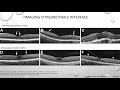
the case was closed under gas tampon here we can see the pre and post of OCD images the best character visual Acuity
was 636 at one month follow-up in our second case we have a 60 year old female with macular hole measuring more
than 400 microns with best corrected visual Equity of 660 on snellen's chart 23 wasp ports were made and [ __ ]
synonym assisted with trick Tommy was performed the ilm was trained with Brilliant Blue and the nasal flap was
peeled foreign a temporal flap was carefully made and
inverted over the macular hole [Music] [Music]
during fluid or exchange the flute was kept nasally to the opted desk like before
foreign here are the pre and post of OCT images the best character visual Acuity was
improved to 636 at one month follow-up in a third case we have a 71 year old female with macular hole less than 400
microns and best corrected visual Equity of 624 23 gauge ports were made and core vitrectomy was done
acetate was used to stay in the vitreous and the vitrectomy was completed the island was then stained with
Brilliant Blue a temporal flap was made which was carefully inverted over the macular hole
[Music] [Music] foreign
was then peaked [Music] fluid air exchange was done and the case
was closed under gas tampon here are the pre and post opposite images the best character visual Acuity
was improved to 612 at one month follow-up inverted ilm flap provides a more
regular needles for fibroblast proliferation which AIDS in macular hole closure nasal LM peeling provides an
additional Advantage by relieving the tangential traction like the conventional technique
thus this modified ilmpl method gives benefit of both the techniques for better closure of large holes thank you
foreign [Music]
Heads up!
This summary and transcript were automatically generated using AI with the Free YouTube Transcript Summary Tool by LunaNotes.
Generate a summary for freeRelated Summaries
Inzicht in Posterior Vitreous Detachment en Epiretinaal Membraan
Deze uitgebreide samenvatting behandelt de stadia van posterior vitreous detachment (PVD), de normale veroudering van het glasvocht, en complicaties zoals vitriomaculaire tractie en epiretinaal membraan. Daarnaast wordt ingegaan op maculagatvorming, de diagnostiek met OCT en fundoscopie, en vasculaire oogafwijkingen zoals CRVO en BRVO, met praktische tips voor herkenning en vervolg.

Detailed Visual Pathway Anatomy and Associated Visual Deficits Explained
Explore the comprehensive anatomy of the visual pathway, from the retina through the optic nerve to the visual cortex, including detailed discussion on fiber crossings, optic radiations, and clinical deficits such as hemianopia and quadrant anopsia. Understand how lesions at different points affect visual fields and learn key mnemonics for retention.

The Evolution and Application of Segmental Spinal Anesthesia
This presentation explores the advancements in segmental spinal anesthesia since its inception in 2006, highlighting its benefits, techniques, and applications in various surgical procedures. The speaker discusses the anatomical considerations, safety concerns, and the effectiveness of different anesthetic agents used in segmental spinal anesthesia.

Comprehensive Insights into Cardiac Hemodynamic Tracings and Heart Failure
Explore detailed analysis of atrial, ventricular, and arterial pressure tracings within the cardiac cycle. Learn to distinguish waveform morphologies, understand pathologic patterns in conditions like constrictive pericarditis and tricuspid regurgitation, and grasp the implications for heart failure diagnosis and management.

Comprehensive ECG Guide to Chamber Enlargement and Hypertrophy Diagnosis
This detailed session explains how to identify right and left atrial abnormalities, ventricular hypertrophy, and dilatation using ECG. Learn key ECG criteria, wave morphology, axis deviations, and diagnostic indices for accurate cardiac chamber enlargement assessment.
Most Viewed Summaries

A Comprehensive Guide to Using Stable Diffusion Forge UI
Explore the Stable Diffusion Forge UI, customizable settings, models, and more to enhance your image generation experience.

Kolonyalismo at Imperyalismo: Ang Kasaysayan ng Pagsakop sa Pilipinas
Tuklasin ang kasaysayan ng kolonyalismo at imperyalismo sa Pilipinas sa pamamagitan ni Ferdinand Magellan.

Mastering Inpainting with Stable Diffusion: Fix Mistakes and Enhance Your Images
Learn to fix mistakes and enhance images with Stable Diffusion's inpainting features effectively.

Pamamaraan at Patakarang Kolonyal ng mga Espanyol sa Pilipinas
Tuklasin ang mga pamamaraan at patakaran ng mga Espanyol sa Pilipinas, at ang epekto nito sa mga Pilipino.

How to Install and Configure Forge: A New Stable Diffusion Web UI
Learn to install and configure the new Forge web UI for Stable Diffusion, with tips on models and settings.

If you found this summary useful, consider buying us a coffee. It would help us a lot!