Introduction to Gastrulation
Gastrulation is a critical phase in human embryonic development occurring at the start of the third week. It transforms the bilaminar germ disc, composed of the epiblast and hypoblast layers, into a trilaminar structure essential for organogenesis. For a deeper understanding of the initial stages, see Comprehensive Guide to Cleavage in Early Human Embryonic Development.
Early Embryonic Structure
- The bilaminar germ disc consists of:
- Epiblast: The upper layer of cells.
- Hypoblast: The lower layer of cells.
- Surrounding cavities include the amniotic cavity (above) and the yolk sac (below).
- The entire structure is suspended within the chorionic cavity, connected via the connecting stalk.
Formation of the Primitive Streak and Axis Determination
- Epiblast cells secrete hyaluronic acid, creating a fluid-filled space and a blotted appearance.
- Chemoattractants stimulate epiblast proliferation at the periphery and migration towards the midline.
- A streak of cells forms at the caudal midline called the primitive streak, extending cranially to form the primitive node (Hensen's node).
- The primitive streak defines the caudal (tail) end; the opposite is the cranial (head) end.
- This process establishes the embryo's body axes (right and left, cranial and caudal).
- The hypoblast cells at the cranial end inhibit primitive streak formation there, aiding head formation by acting as the anterior visceral endoderm.
Cellular Changes During Gastrulation
- Epiblast cells lose adhesion molecules like E-cadherin to detach and invaginate through the primitive streak.
- These cells undergo an epithelial-to-mesenchymal transition (EMT), changing shape into bottle/flask cells and then mesenchymal cells with irregular shapes and motility.
Formation of Germ Layers
- Invaginating epiblast cells form:
- Definitive endoderm: Mesenchymal cells displace hypoblast cells, which are relocated to the yolk sac.
- Mesoderm: Migrating cells populate the space between ectoderm and endoderm.
- Ectoderm: Remaining epiblast cells form the outer layer.
Primitive Groove and Primitive Pit
- The depression formed by invaginating cells is the primitive groove.
- The groove extends into the primitive pit, where further invaginations contribute to the formation of the notochord, a rod-like mesodermal structure crucial for signaling future development.
Differentiation and Clinical Significance
- The displaced hypoblast stimulates extraembryonic mesoderm to form blood islands, precursors to blood cells.
- The entire process leads to the formation of the trilaminar germ disc: ectoderm, mesoderm, and endoderm.
- Persistence of the primitive streak beyond the fourth week can cause sacrococcygeal teratoma, a tumor containing diverse tissue types like hair, teeth, and nails due to the pluripotent nature of primitive streak cells.
Why the Term 'Gastrulation'? Explanation and Historical Context
- The term derives from early studies in simpler organisms (e.g., sea urchins) where gastrulation involved formation of a primitive gut (archenteron).
- In humans, the term persists even though primitive gut formation is a later event.
- This is a convention reflecting embryological history rather than direct gut formation at this stage.
Summary
- Epiblast cells proliferate and migrate to form the primitive streak.
- Loss of cell adhesion facilitates migration and transformation into mesenchymal cells.
- Subsequent formation of three germ layers, ectoderm, mesoderm, endoderm, occurs through coordinated cell movements and differentiation.
- The primitive streak is essential for establishing body axes and germ layer formation.
- Understanding these steps provides insight into normal development and congenital anomalies.
This lecture provides a foundational understanding of gastrulation, an indispensable process shaping the embryonic body plan and germ layer formation in humans. To appreciate the stages following gastrulation, particularly blastocyst embedding and implantation, consider reviewing the Comprehensive Guide to Human Blastocyst Implantation Process. For insights into early cardiac development which follows gastrulation, see Comprehensive Guide to Heart Development: From Heart Tube to Valves. Finally, to better understand fertilization that precedes these stages, refer to the Comprehensive Guide to Fertilization: Process, Steps, and Effects.
Hello! I am Dr Aizaz from medicovisual.com and in this visual lecture we will talk about gastrulation. Here is the embryo at the start of third week of development. If we cut open this embryo, if we divide the embryo with a knife into two parts
and if we remove this part of the embryo and we observe this cross section of the embryo what we will see is that it consists of two types of cell forming the bilaminar germ disc the upper layer of cell is called epiblast while the lower layer of cells is called hypoblast.
Amniotic cavity surrounds this embryo from above while the secondary or definitive yolk sac surrounds this embryo from below. Then this yolk sac membrane or yolk sac endoderm, it surrounds this yolk sac. Of course,
then there is extra embryonic mesoderm and whole this structure it is floating freely within this chorionic cavity but it is still connected with the connecting stalk and of course all this structure is surrounded by cytotrophoblast cells and of course outside this will be the syncytio-
trophoblast which has not been shown in this diagram. Let's focus on this bilaminar germ disc. If we undo the cut here we will see that it is a disc like structure consisting of hypoblast as well as epiblast layers and this is called bilaminar germ disc. Let's focus on this.
So, here is this bi- laminar germ disc and let's make a copy of this bilaminar germ disc and in this copy version let's cut it from here. So, if we cut it from here and we observe it from front for example you are standing here and you are observing it from front how it will look like?
So, here is this bilaminar germ disc from front. Again it consists of epiblast layer as well as hypoblast layer. Now at this stage what happens and of course we are talking about third week of development so at the start of third week of development what will happen
is that this epiblast layer they will release some special substances in between these two layers, in between the hypo and epiblast layer. What is this substance? It will release lots of hyaluronic acid in between these two layers. This hyaluronic acid is a hygroscopic compound
it can bind with and it can pick up lots of water molecules and thus a fluid filled space will be formed in between hypo and epiblast cell layer and in this way we will get a blotted appearance of the embryo. Here we can see that we have this blotted up appearance of the embryo
then these midline epiblast cells they will release some chemo attractants and what they and what these chemo attractants will do is that they will cause growth of epiblast in the periphery. Now what these chemoattractants will do is that they will cause proliferation of epiblast in the
periphery and they will attract this epiblast this newly formed epiblast towards the midline. So, as these newly formed epiblast aggregate in the midline what we will see is that in the caudal midline there will be formation of a streak of cells, it will start
in the mid of caudal midline and it will extend caudally as well as it will extend cranially and at the cranial end it will form the swelling or node called primitive node and this streak of cell it is called primitive streak. So, here is this structure called primitive streak on
the cut section as well as while we view it from the top and this node is called primitive node or Hansen’s node. Remember that this structure is formed only at caudal end. Caudal end means tail end. Of course, we humans do not have tail but still traditionally embryologists
call it caudal end and this end as cranial end. So, with the formation of primitive streak we can clearly say that this end is caudal end on which the primitive streak is formed while the other end that is opposite to primitive streak is the cranial end.
Before the formation of primitive streak just by looking at this bilaminar germ disc we cannot tell that which end is the cranial and which end is the caudal end. Along with that in the midline as this primitive streak is formed we can say that this is the right side of the embryo and this is the left
side of the embryo. So, axis determination is one of the important function of the primitive streak. Now here an important question arises that why primitive streak only forms at the caudal end? What stops the formation of primitive streak at the cranial end?
Actually, we have these hypoblast cells and these hypoblast cells they are like wise old man, they give important instructions to the overlying hypoblast cells, the hypoblast cells of the cranial end inhibit the formation of primitive streak
at the cranial end of epiblast. As they do so the epiblast in the caudal end they quickly forms the primitive streak as they are so keen to do so. This cranial end hypoblast also helps in formation of head structure so it is called anterior
visceral endoderm. Anterior? *** the anterior here? Of course, this is the cranial end this is not the anterior and so why we call it anterior visceral endoderm? I think it should be called cranial visceral endoderm rather than anterior visceral endoderm. Actually, this name is given
to this structure by the mouse embryologist and in mouse this structure is not just cranial but it is also anterior. Well! let's see how? So, here is a mouse and it has to pass through this door the first structure of his body that will cross the door is its head so it is called anterior
I mean the head of embryo is anterior, anterior means something that comes before the other part and the last part of his body to cross that this door is his tail so his caudal end or his tail is also the posterior end. So, in mouse anterior as well as the cranial both of these terms are
synonymous. Similarly, the term caudal as well as posterior both of these terms are synonymous. But this does not happen with humans. Because let's say if this human has to cross this door both the head as well as, of course humans
do not have tail, both the head as well as his buttocks or hips both of them will cross the door at the same time so we cannot demarcate the head and hips in terms of anterior and posterior. So, in humans the head is cranial but it is not necessarily anterior [Music]
but still conventionally we call it anterior visceral endoderm and you have to do the same. Right, epiblast cells just like any other type of epithelial cells they are closely bound with each other kind of like holding hands of each other and that is due to special proteins called e-cadherin
proteins that keep them formed together with each other. Now what happens that as these primitive streak cells come into the midline, in the centre these midline epiblast cells what they do is that they don't regulate the e-cadherin thus they lose their hands and they are no more bond
with each other. So, what happens here in this strange land of primitive seek these epiblasts, these midline epiblast cells what they do is that first they invite these guests into this area and then they remove their hands and of course, now what will happen that they will not be able to
stay together. So, let's suppose that this midline epiblast cell it loses its e-cadherin molecule so it will tend to fall into this space and as it turns so it loses its particular shape and it forms a bottle like shape a flask like shape and now it is called bottle cell or flask cell.
So, ultimately it will fall into this space and now we call it mesenchymal cell. This is the epithelial cell and this is a mesenchymal cell. The difference between epithelial cell and mesenchymal cells is that number one the mesenchymal cell it has an irregular shape
but epithelial cell it has a particular shape. For example, this epiblast cell these are, these epiblast cells they are columnar in shape but this mesenchymal cell it has some irregular shape. Secondly, the epithelial cells they are tightly bound together. They are closely packed together
with each other but that is not true for mesenchymal cells. So, what we can say is that as this epiblast it invaginated inward into this space what happened with this epiblast cell is that it underwent epithelium to mesenchymal transition. So, as more and more cell invaginate
inward from this point here we will see that a depression, a small depression is formed here. Here you can see there is a depression the name of this depression is primitive groove. So, more and more cells will come into this space and here the structure that
is formed is mesenchyme. Mesenchyme is basically the embryonic connective tissue. It consists of ground substance that is filled up with hyaluronic acid and other substances and within this ground substance mesenchymal cells are loosely arranged.
On this 3D diagram you can see that this was the primitive streak and as the cell invaginated inward a groove is formed in the, in the centre of this primitive streak so this groove is named as primitive groove. Here on the cut section you can see this
primitive groove and on the 3D diagram you can also see the primitive groove. This primitive groove it extends caudally into an other groove, a shallow depression that is called primitive pit. So, that there is that primitive pit.
Now how this primitive pit forms? What happens that here also some cells invaginate inward, right, some cells invaginate inward and they move forward in between the space, between epi and hypoblast and in this way this rod-like structure will be formed here below
this epiblast layer this rod like structure is called notochord. We will discuss that how it form and what is its function in the next lecture. Right, and regarding the primitive groove as you know that how it forms? The cell invaginate here from this groove the cell invaginates inward
and this leads to formation of this depression called primitive groove. Right? So, now we have three layers this is the epiblast layer, this is the hypoblast layer and in between them there is another layer of mesenchymal cells. And now we have this three layers. Is
this the process called gastrulation? Well! No, it is not that simple unfortunately. What happens here is that as the embryo is growing in size the simple diffusion process is not simply enough to meet the nutritious need of the embryo. So, this is the right time that blood cells should form.
As you know that here we have this yolk sac endoderm or yolk sac membrane and outside this yolk sac endoderm is extra embryonic splanchnopleuric mesoderm. Now these mesenchymal cells they leave this space
and they come here into the place of hypoblast and they displace these hypoblast into the yolk sac endoderm. So, hypoblasts are displaced to yolk sac endoderm and here these mesenchymal cells come. So, these mesenchymal cell will almost completely replace the hypoblast and those hypoblasts they
will then be displaced to the yolk sac endoderm. These mesenchymal cells as they come here they will not remain mesenchymal they will then again undergo mesenchymal two epithelial transition and here they will form the definitive endoderm. They will acquire the hypoblast
like shape. Of course, they are not hypoblast. They will acquire hypoblast like shape and they will form the definitive endoderm. What about the hypoblast that were displaced here into the yolk sac endoderm? As I have told you the hypoblast they are like wise old men. They have
a real bossy nature. What they will do is that they will order the overlying extra embryonic mesoderm that “Heyy! extra embryonic mesoderm start creating blood island.” And of course, it will do so. The blood island will be formed in the extra embryonic splanchnopleuric mesoderm
on the order of yolk sac endoderm. In books what they say is that the underlying yolk sac endoderm induces the formation of blood island into the extra embryonic splanchnopleuric mesoderm. What is the function of blood island? For now just remember that it helps in formation of blood cells
we will discuss the details of blood island in upcoming lectures. So, as these mesenchymal cells that were lying here they displaced the hypoblast and came here and differentiated into the definitive endodermal cell this space is now empty so more
epiblast cells will proliferate and invaginate inward. They will migrate towards the midline and invaginate inward through the primitive groove and again they will come into this space to form the definitive mesoderm. The definitive mesenchyme will be formed here.
So, now ultimately we have three germ layers due to the faithful work done by the primitive streak. As the primitive streak has performed its function nicely during the fourth week of development primitive streak usually disappears
but unfortunately sometimes the primitive streak failed to disappear and the remnants of primitive streak remains and it forms what we call as sacrococcygeal teratoma. here is a new born with a sacrococcygeal teratoma in the hip region here this is
called Teratoma. Teratoma means monstrous tumor because it looks like a monster. As you know that these epiblast cells and this primitive streak it is the precursor of all different types of cells it forms the three germ layers that are ultimately
going to form the complete embryo. So, this tumor it consists of all different varieties of cell. There may be nail, there may be teeth and hair and all those types of adult cells. So, it may look like a monster so that is why we call it teratoma, the monstrous tumor.
So, now here we have three germ layers the outer layer is called ectoderm, the inner layer is endoderm and in between them is the mesoderm. Meso means in the middle, that comes in the middle. And it consists of mesenchymal tissue. This is trilaminar germ disc and the origin of all these
germ layers is epiblast cells. This process of formation of trilaminar germ disc is termed as gastrulation. Gastrulation? There is no formation of stomach here, there is no formation of gut tube here why the hell we call it gastrulation? Well! this is confusing so let me explain why it
is called gastrulation? Actually, this process was first discovered in small animals for example, sea urchins and this primitive organism their blastula it consists of a hollow spherical ball of cell and during the process of gastrulation, their process of gastrulation is very simple it looks
like that someone has pinched here with a thumb. Right, so someone has pinched here with the thumb. So, here they will invaginate inward as you can see here that here it has invaginated inward and in this way what happens is that here this part here, here we have formation of primitive gut or
Archenteron. The water and food material it enters first into this primitive gut and it is released outside through this blastopore. So, during gastrulation what is happening here is that there is formation of primitive gut during the process of gastrulation but along with that we have also
the formation of germ layers. Here is the endoderm that has formed along with that there is ectoderm and in some organism in between them the mesoderm is also formed. So, three germ layers are formed here along with the formation of three germ layers the primitive gut is also formed.
So, scientists initially thought that the formation of primitive gut that is gastrulation is the most important process here so they simply ignored the trilamination. So, rather than calling this process as trilamination they simply call it gastrulation. Why? Because in
small organism primitive gut is formed here but no such process occur in human. In human, during the gastrulation process no gut forms but still somehow it is called gastrulation. Why so? By now you must have understood this. Yes! You got it right. Conventions my friend conventions. Right?
So, finally let's review it. So, first of all this is the epiblast layer what happens that it releases chemo attractants that causes proliferation and migration of epiblast from the periphery into the caudal midline
and then it down regulates the e-cadherin thus these epiblasts they tend to fall into the space. Firstly forming the bottle cells and then they come here forming the mesenchymal cell. Some of these mesenchymal cells they will displace the hypoblast to yolk sac endoderm and
here they will differentiate into the definitive endoderm. The more epiblast will invaginate inward and they will form the mesenchymal cells here that will form the definitive mesoderm and the cells that remain behind they will form the ectoderm. So, ultimately through this process
of gastrulation we have three germ layers the ectoderm, the mesoderm and the endoderm. So, that was about gastrulation. Thank you so much for watching this video.
The primitive streak forms at the caudal midline of the epiblast and serves as the site where epiblast cells detach, migrate, and invaginate to form the germ layers. It establishes the body axes (cranial-caudal, right-left) and is essential for the formation of the trilaminar germ disc, including ectoderm, mesoderm, and endoderm.
Epiblast cells undergo an epithelial-to-mesenchymal transition (EMT), losing adhesion molecules like E-cadherin to detach and migrate through the primitive streak. Some cells displace hypoblast cells to form definitive endoderm, others migrate between layers to become mesoderm, and remaining epiblast cells become ectoderm.
Hypoblast cells at the cranial end act as the anterior visceral endoderm, inhibiting primitive streak formation there. This spatial regulation ensures the primitive streak forms only at the caudal end, helping establish the head-tail (cranial-caudal) axis during embryonic development.
If the primitive streak persists beyond the fourth week, it can give rise to sacrococcygeal teratoma, a tumor that contains various tissue types such as hair, teeth, and nails. This occurs because primitive streak cells are pluripotent and can differentiate into multiple tissues abnormally.
The primitive pit is an invagination at the cranial end of the primitive streak. Cells migrating through this pit give rise to the notochord, a rod-like mesodermal structure that provides critical signaling cues for the development of the neural tube and axial skeleton.
The term 'gastrulation' originates from studies in simpler organisms like sea urchins, where it involves formation of the primitive gut. In humans, although the primitive gut forms later, the term persists to describe this key phase of germ layer formation. It reflects embryological history rather than a direct gut formation event at this stage.
Displaced hypoblast cells are relocated to the yolk sac, where they stimulate the formation of blood islands in the extraembryonic mesoderm, precursors to blood cells. This coordination supports both embryonic germ layer formation and early circulatory system development.
Heads up!
This summary and transcript were automatically generated using AI with the Free YouTube Transcript Summary Tool by LunaNotes.
Generate a summary for freeRelated summaries

Comprehensive Guide to Cleavage in Early Human Embryonic Development
This detailed summary explains the process of cleavage during the first week of human embryonic development, covering key stages from fertilization to blastocyst formation. Learn about the significance of cleavage, stages like morula and blastocyst, and important terms such as trophoblast, embryoblast, and embryonic pole.
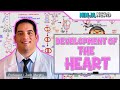
Comprehensive Guide to Heart Development: From Heart Tube to Valves
Explore the detailed process of heart development, from the formation of the heart tube and pericardial cavity to cardiac looping and valve formation. Understand key structures like the septa, atrioventricular canals, and outflow tracts, and learn how neural crest cells and growth factors influence heart morphogenesis.

Comprehensive Guide to Human Blastocyst Implantation Process
This detailed summary explains the stages of blastocyst implantation in the uterine endometrium, including the role of the zona pellucida, decidual reaction, and trophoblastic invasion. Learn about the timing, site, types, and clinical significance of implantation with clear, exam-focused insights.
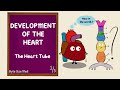
Heart Development Explained: From Heart Tube to Cardiac Looping
This video details the early stages of heart development, focusing on the formation and transformation of the heart tube from the mesoderm. It explains embryonic folding, heart tube structure, and the process of cardiac looping that shapes the four-chambered heart.
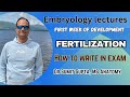
Comprehensive Guide to Fertilization: Process, Steps, and Effects
This lecture provides a detailed overview of fertilization, covering the definition, site, prerequisites like capacitation and acrosomal reaction, the four key steps, and the critical role of the calcium wave. Learn how fertilization leads to zygote formation and prevents polyspermy, with clear explanations and exam tips.
Most viewed summaries

A Comprehensive Guide to Using Stable Diffusion Forge UI
Explore the Stable Diffusion Forge UI, customizable settings, models, and more to enhance your image generation experience.

Kolonyalismo at Imperyalismo: Ang Kasaysayan ng Pagsakop sa Pilipinas
Tuklasin ang kasaysayan ng kolonyalismo at imperyalismo sa Pilipinas sa pamamagitan ni Ferdinand Magellan.

Mastering Inpainting with Stable Diffusion: Fix Mistakes and Enhance Your Images
Learn to fix mistakes and enhance images with Stable Diffusion's inpainting features effectively.

Pamamaraan at Patakarang Kolonyal ng mga Espanyol sa Pilipinas
Tuklasin ang mga pamamaraan at patakaran ng mga Espanyol sa Pilipinas, at ang epekto nito sa mga Pilipino.

How to Install and Configure Forge: A New Stable Diffusion Web UI
Learn to install and configure the new Forge web UI for Stable Diffusion, with tips on models and settings.

If you found this summary useful, consider buying us a coffee. It would help us a lot!