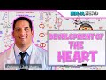
Introduction to Heart Development
- The heart develops from the mesoderm, one of the three germ layers (ectoderm, endoderm, mesoderm).
- The lateral plate mesoderm splits into somatic and splanchnic layers; the heart originates in the splanchnic layer.
- Endoderm stimulates mesodermal cells to form blood islands, initiating vasculogenesis and forming paired heart tubes.
Embryonic Folding and Heart Tube Formation
- The embryo undergoes lateral and cephalocaudal folding:
- Lateral folding brings the paired heart tubes together, forming a single heart tube within the pericardial cavity.
- Cephalocaudal folding moves the heart from the cranial (head) region to the thoracic region.
- Folding is essential to position the heart correctly and integrate it into the developing embryo.
Structure of the Heart Tube
- The heart tube has a venous end (sinus venosus) and an arterial end (truncus arteriosus).
- Layers of the heart tube:
- Endocardium: endothelial lining.
- Myocardium: cardiac muscle layer formed by cardiac myoblasts.
- Cardiac jelly: specialized connective tissue between endocardium and myocardium.
- Epicardium: outer layer formed by migrating mesothelial cells.
Overview of Mature Heart Structures
- Four chambers: right atrium, left atrium, right ventricle, left ventricle.
- Atria have smooth posterior walls (venous openings) and rough anterior walls with pectinate muscles.
- Ventricles have trabeculae carneae (muscular ridges) in inflow portions and smooth outflow tracts.
- Valves:
- Atrioventricular valves: tricuspid (right), mitral (left) with chordae tendineae and papillary muscles.
- Semilunar valves: pulmonary (right), aortic (left).
Heart Tube Segments and Their Derivatives
- Truncus arteriosus: forms ascending aorta and pulmonary trunk; separated by spiral aorticopulmonary septum.
- Bulbus cordis (divided into distal third - truncus arteriosus, middle third - conus cordis, proximal third):
- Conus cordis forms smooth outflow tracts (infundibulum and aortic vestibule).
- Proximal third forms trabeculated right ventricle.
- Primitive ventricle: forms trabeculated left ventricle.
- Primitive atrium: forms rough anterior walls of both atria.
- Sinus venosus: venous inflow, connects to primitive atrium via sinoatrial orifice.
Blood Flow Through the Heart Tube
- Blood enters from sinus venosus into primitive atrium.
- Passes to primitive ventricle.
- Moves through bulbus cordis.
- Exits via truncus arteriosus into arterial system.
Cardiac Looping
- The heart tube undergoes looping to form the shape of the mature heart.
- The truncus arteriosus, bulbus cordis, and primitive ventricle bend ventrally and shift forward and downward.
- Primitive atrium and sinus venosus shift dorsally and upward, positioning behind the ventricles.
- This rearrangement aligns the inflow and outflow tracts properly.
Summary
- The heart develops from paired tubes in the mesoderm that fuse and fold to form a single heart tube.
- Embryonic folding and cardiac looping are critical for proper heart positioning and chamber formation.
- The heart tube segments correspond to specific adult heart structures.
- Understanding these early stages is essential for grasping congenital heart development.
For further details on atrial development, stay tuned for part two of this series.
For a deeper understanding of heart development, check out our Comprehensive Guide to Heart Development: From Heart Tube to Valves and Understanding Human Physiology: A Comprehensive Overview of the Circulatory System. Additionally, you may find the Comprehensive Guide to Heart Conduction and ECG Fundamentals helpful for insights into the electrical aspects of heart function.
Hello welcome to Byte Size Med. This video is on the development of the heart and part one is on the heart tube. The heart develops from the mesoderm. Remember the three germ layers? There's
the ectoderm, the endoderm and the mesoderm. The mesoderm has three parts. The paraxial mesoderm, the intermediate mesoderm and the lateral plate mesoderm. The lateral plate mesoderm develops cavities that fuse to form an intra-embryonic cavity.
That splits the lateral plate mesoderm into two layers, the somatic layer and the splanchnic layer . Now the heart develops in the splanchnic layer of the lateral plate mesoderm. It starts off as a cluster of cells. The endoderm underneath stimulates these cells to differentiate into
blood Islands. These blood Islands form vessels and that process is called vasculogenesis. The vessel that they form here is called the heart tube and there are two. So paired heart tubes. If we go back to those three germ layers, these are the two heart tubes. One on each side.
The heart sits in the pericardial cavity. Now that space forms from the intra-embryonic cavity. If you look at these sections as best as I could draw them you'll notice that the sides are coming together. That's because the embryo is folding. I'm going to try and simplify what that is.
The hard part about embryology is the fact that so many things happen at once and things keep changing. That's why there are so many sections at so many different angles . Assume that this is the embryo now we're looking at it from above and we've removed the amnion.
This is the head end of the embryo or the cranial end or the cephalic end and this is the tail end or the caudal end. This would be the dorsal surface of the embryo. Now the sections we saw earlier, they were across like this at different ages of the embryo.
Transverse sections or cross sections. So you can see the midline, the sides and the layers. A section going this way would be a longitudinal section or a sagittal section. The embryo folds during its development in two directions, lateral and
cephalocaudal. Lateral folding would be the sides coming together closing the ventral body wall. Cephalocaudal folding would be the bending of the head and the tail ends. To see lateral folding, we're gonna have to use transverse sections and when the embryo folds,
the two heart tubes come together and unite to form one heart tube in the pericardial cavity. Now in a section like this, you'll also see the dorsal aortae which continue from the heart tube but ignore those for now. So the lateral folding results in a single heart tube.
But the embryo folds cephalocaudally as well and for that we're going to use a sagittal section which would look something like this.This is the cephalic end and this is the caudal end. Now the fascinating thing about heart development is the fact that the heart starts developing at the cephalic end, in the
head making things just as confusing as two heart tubes. But the folding is how it gets sorted out. Cephalocaudal folding. So when the head end folds, the heart comes towards the thoracic region. Initially the heart tube and the pericardial coelom are separate and their relationship
changes as it folds. Finally the heart tube gets pushed into the pericardial cavity. Now we'll pick up that heart tube and reorient it to look at it vertically. It has a venous end and an arterial end.
The heart tube was a vessel with endothelium, that endothelial lining forms the endocardium. It was surrounded by cardiac myoblasts and they form the myocardial layer. The myocardium produces a specialized connective tissue which separates the two layers this is called the cardiac jelly.
The outer epicardial layer which is the visceral layer of the pericardium is formed by migration of mesothelial cells which then cover the myocardium. And these are the layers of the heart tube. It still looks nothing like what the heart is supposed to look like and it's incredible how
it goes from this to that. But to understand how let's first look at some of the structures of the heart that need to develop and we'll come back here. The heart has four chambers, the right atrium, the left atrium, the right ventricle and the left ventricle. First let's look at the Atria. The
right atrium receives blood from the superior vena cava, the inferior vena cava and the coronary sinus. The left atrium receives blood from the four pulmonary veins. In both the atria, all these openings are on the posterior wall making it smooth. So they have a smooth posterior wall.
The anterior wall on the other hand is rough, with muscular ridges called muscle pectinati. And those are the two parts of the atria. Between the atria and the ventricles are the atrioventricular valves, the tricuspid valve on the right and the mitral valve on the left.
The free ends of the valve leaflets are attached to chordae tendinae, which in turn are attached to papillary muscles on the wall of the ventricles. The walls of the ventricles have muscular ridges and bridges called trabeculae carneae.
That makes them rough or trabeculated. This is the rough inflow portion of the ventricles. The right ventricle pumps blood into the pulmonary trunk and the left ventricle into the ascending aorta. The outflow tracts that lead into these vessels are smooth.
The outflow tract of the right ventricle is called the infundibulum or the conus arteriosus, while that of the left ventricle is called the aortic vestibule. These are the smooth outflow tracts of the ventricles. Between the ventricles and those big vessels are the semilunar valves.
That's the pulmonary valve on the right and the aortic valve on the left. The ventricles pump blood into the ascending aorta and the pulmonary trunk. And now let's go back to the heart tube. It has alternating dilations and constrictions which are going to form the different chambers
of the heart. At the top here we have the arterial end which is going to lead into the aortic Sac with the aortic roots. The first part of the heart tube is called the truncus arteriosus. Below that is the bulbus cordis and I'm going to divide that which I'll explain in a bit. Next
we have the Primitive ventricle and then the Primitive atrium. The venous end is the sinus venosus. Starting at the top, the truncus arteriosus which sounds like an arterial trunk forms the ascending aorta and the pulmonary trunk. Now these two will separate when a spiral aorticopulmonary
septum forms between them. The bulbus cordis forms the smooth outflow tracts of the ventricles, that's the infundibulum on the right and the aortic vestibule on the left. But the proximal third of the bulbus cordis forms the right ventricle. The rough trabeculated part of the right ventricle.
The bulbus cordis actually has three parts, a distal third, a middle third and a proximal third. The distal third is the truncus arteriosus, the middle third is also called the conus cordis and if you remember that the smooth outflow tract of the right ventricle is also called the conus
arteriosus, you'll remember that the outflow tracts of both the ventricles comes from the conus cordis. The proximal third doesn't have a special name, but it forms the trabeculated part of the right ventricle. The trabeculated part of the left ventricle is formed from the Primitive ventricle.
Between the bulbus cordis and the Primitive ventricle is a bulboventricular sulcus. This is the site of the interventricular foramen, because it's between the developing right ventricle and the developing left ventricle. This will be the site of formation of the
interventricular septum, which will eventually separate the two chambers. The Primitive Atrium forms the two atria, the rough anterior walls of both the Atria. Between the developing atria and the developing ventricles is the atrioventricular canal
which won't close, but it will split to separate the right and the left side. What's left in the atria is the smooth posterior walls which I'll explain when I talk about atrial development because that's when I'll talk about this sinus venosus but between the sinus venosus
and the Primitive Atrium there is a sinoatrial orifice. Now let's see the path that blood takes through the heart tube. It starts at the venous end from the sinus venosus into the Primitive Atrium. Let's put those two together as the Atria and their inflow tracts. Next up on the path is the
Primitive ventricle, the bulbus cordis and the truncus arteriosus, which we'll put together as the ventricles and their outflow tracts. Blood then gets pumped out of the arterial end. Now this heart tube has to go from looking like this to this and that happens by a process
called looping, Cardiac looping. And now you'll see why I grouped them. The truncus arteriosus, the bulbus cordis and the Primitive ventricle. They bend ventrally. The ventricles enlarge and this part of the heart tube comes forward and shifts lower down. Meanwhile the Primitive Atrium and
the sinus venosis shift backward, so dorsally and upwards so that they come behind the ventricles. Now in front we have the truncus arteriosus, the conus cordis and the proximal third of the bulbus cordis The Primitive ventricle and behind, we have the Primitive atrium.
If we flip it over to look at the back the sinus venosus opens into the Primitive Atrium through that sinoatrial orifice. And now it's starting to look more like the heart on the outside.
The truncus arteriosus forms the ascending aorta and the pulmonary trunk. The conus cordis forms the smooth outflow tracts of both the ventricles. The proximal third of the bulbus cordis forms the trabeculated part of the right ventricle. The Primitive ventricle forms the
trabeculated part of the left ventricle and the Primitive Atrium forms the rough parts of both the right and the left Atria. In part two, we'll look at how these two atria develop. But all that was the heart tube. I hope this video was helpful, if it was you can give
it a like and subscribe to my channel. Thanks for watching and I'll see you in the next one! :)
Heart development begins with the formation of paired heart tubes from the mesoderm, which then fuse to create a single heart tube. This tube undergoes embryonic folding and cardiac looping, essential processes that position the heart correctly and shape it into its mature form, including the formation of four chambers.
Embryonic folding involves lateral and cephalocaudal movements that bring the paired heart tubes together into a single heart tube located in the thoracic region. This folding is crucial for integrating the heart into the developing embryo and ensuring proper alignment of the heart's structures.
The heart tube consists of several layers: the endocardium (inner lining), myocardium (muscle layer), cardiac jelly (connective tissue), and epicardium (outer layer). Each layer plays a vital role in the heart's function, with the myocardium responsible for contraction and blood pumping.
The heart tube has several segments, including the truncus arteriosus (which forms the ascending aorta and pulmonary trunk), bulbus cordis (which develops into parts of the ventricles), and the primitive atrium (which becomes the atria). Each segment is crucial for the formation of specific adult heart structures.
Blood flows into the heart tube starting from the sinus venosus into the primitive atrium, then to the primitive ventricle, through the bulbus cordis, and finally exits via the truncus arteriosus into the arterial system. This flow is essential for the heart's function during development.
Cardiac looping is the process where the heart tube bends and rearranges to form the shape of the mature heart. This process is vital for aligning the inflow and outflow tracts, ensuring that blood flows correctly through the heart's chambers.
For more detailed insights into heart development, you can explore our Comprehensive Guide to Heart Development: From Heart Tube to Valves and Understanding Human Physiology: A Comprehensive Overview of the Circulatory System. Additionally, the Comprehensive Guide to Heart Conduction and ECG Fundamentals offers valuable information on the electrical aspects of heart function.
Heads up!
This summary and transcript were automatically generated using AI with the Free YouTube Transcript Summary Tool by LunaNotes.
Generate a summary for freeRelated Summaries
Comprehensive Guide to Heart Development: From Heart Tube to Valves
Explore the detailed process of heart development, from the formation of the heart tube and pericardial cavity to cardiac looping and valve formation. Understand key structures like the septa, atrioventricular canals, and outflow tracts, and learn how neural crest cells and growth factors influence heart morphogenesis.
Comprehensive Guide to Cardiac Electrophysiology and Heart Conduction System
Explore the intrinsic electrical properties of the heart, including automaticity, nodal and contractile cells, and the detailed conduction pathway from the SA node through the Purkinje fibers. Understand how action potentials are generated and propagated, and how ion channels contribute to cardiac muscle contraction and relaxation.

Comprehensive Heart Anatomy, Physiology, and Electrolyte Balance Explained
This detailed lecture covers heart anatomy, muscle types, electrophysiology, and critical electrolyte imbalances. Learn how cardiac muscle functions, the role of ions in heartbeats, and the clinical significance of electrolyte disorders.

Comprehensive Guide to Heart Conduction and ECG Fundamentals
Explore the detailed physiology of the heart's conduction system, including pacemaker activity, cardiac muscle properties, and the generation of ECG waveforms. Understand how electrical impulses travel through the heart to coordinate contraction and how this relates to ECG interpretation.

Understanding Human Physiology: A Comprehensive Overview of the Circulatory System
This video provides an in-depth exploration of human physiology, focusing on the circulatory system. It covers essential components such as blood, blood vessels, and the heart, while also discussing the differences between various circulatory systems and their functions. The session aims to equip viewers with a complete understanding of the chapter's key concepts and potential exam questions.
Most Viewed Summaries

A Comprehensive Guide to Using Stable Diffusion Forge UI
Explore the Stable Diffusion Forge UI, customizable settings, models, and more to enhance your image generation experience.

Kolonyalismo at Imperyalismo: Ang Kasaysayan ng Pagsakop sa Pilipinas
Tuklasin ang kasaysayan ng kolonyalismo at imperyalismo sa Pilipinas sa pamamagitan ni Ferdinand Magellan.

Mastering Inpainting with Stable Diffusion: Fix Mistakes and Enhance Your Images
Learn to fix mistakes and enhance images with Stable Diffusion's inpainting features effectively.

Pamamaraan at Patakarang Kolonyal ng mga Espanyol sa Pilipinas
Tuklasin ang mga pamamaraan at patakaran ng mga Espanyol sa Pilipinas, at ang epekto nito sa mga Pilipino.

How to Install and Configure Forge: A New Stable Diffusion Web UI
Learn to install and configure the new Forge web UI for Stable Diffusion, with tips on models and settings.

If you found this summary useful, consider buying us a coffee. It would help us a lot!