Introduction to Heart Development
This video provides an in-depth explanation of the embryonic development of the heart, starting from the formation of the heart tube to the complex septation and valve formation processes.
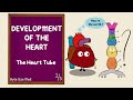
Early Heart Formation
- The heart begins as two heart tubes derived from the splanchnic layer of the lateral plate mesoderm.
- The endoderm secretes vascular endothelial growth factor (VEGF), stimulating differentiation of mesodermal cells into angioblasts (forming blood vessels and endocardium) and hemocytoblasts (forming blood cells).
- Lateral folding of the embryo fuses the two heart tubes into a single heart tube within the pericardial cavity.
- The heart tube is suspended by the dorsal mesocardium.
Heart Tube Structure
- The heart tube consists of three layers:
- Endocardium (inner endothelial layer)
- Myocardium (outer cardiac muscle layer)
- Cardiac jelly (connective tissue between endocardium and myocardium)
Sagittal and Cross-Sectional Views
- Sagittal folding moves the heart tube from the cranial region into the thorax.
- Cross-sectional views highlight the mesodermal layers and the formation of the pericardial cavity.
Segmentation of the Heart Tube
- The heart tube is divided into key regions:
- Truncus arteriosus (forms pulmonary artery and aorta)
- Bulbus cordis (forms right ventricle and outflow tracts)
- Primitive ventricle (forms left ventricle)
- Primitive atria (forms left and right atria)
- Sinus venosus (inflow tract receiving venous blood)
Cardiac Looping
- Cardiac looping repositions the heart tube to the left side of the midline.
- Dynein proteins are essential for proper looping; defects can cause dextrocardia or situs inversus.
- The primitive atria move posteriorly and superiorly, overlaying the ventricles.
Formation of Atrioventricular (AV) Canals and Valves
- Neural crest cells migrate to form endocardial cushions in the AV canal region.
- Fusion of anterior and posterior cushions creates the septum intermedium, dividing the AV canal into right and left canals.
- Valve leaflets, annulus rings, and chordae tendineae develop from these cushions, forming the mitral and tricuspid valves.
Atrial Septation
- The septum primum grows downward toward the septum intermedium, initially leaving the ostium primum.
- After closure, the ostium secundum forms in the septum primum.
- The septum secundum develops to partially cover the ostium secundum, creating the foramen ovale.
- The foramen ovale allows fetal blood to bypass the lungs; failure to close postnatally results in a patent foramen ovale.
Ventricular Septation
- The muscular interventricular septum grows upward from the apex.
- The membranous interventricular septum forms from the septum intermedium and fuses with the muscular septum.
- Failure of fusion causes ventricular septal defects.
Development of Inflow Tracts
- The sinus venosus and its horns receive blood from common cardinal, umbilical, and vitelline veins.
- The left horn regresses to form the coronary sinus.
- The right horn contributes to the formation of the superior and inferior vena cava.
Outflow Tract Formation and Septation
- Neural crest cells form truncal and bulbar ridges in the truncus arteriosus and bulbus cordis.
- These ridges fuse and spiral to form the aorticopulmonary septum, separating the aorta and pulmonary trunk.
- The septum's spiral nature ensures proper alignment of outflow tracts with respective ventricles.
Semilunar Valve Formation
- Endocardial cushions develop at the junction of the bulbus cordis and truncus arteriosus.
- Four cushions (anterior, posterior, right, left) form and remodel to create the aortic and pulmonary semilunar valves.
- Rotation of the outflow tract positions the aorta posteriorly and to the right, and the pulmonary trunk anteriorly and to the left.
Summary
This comprehensive overview covers the critical stages of heart development, emphasizing the roles of mesodermal differentiation, folding, looping, septation, and valve formation. Understanding these processes is essential for recognizing congenital heart defects and their embryological origins.
For a deeper understanding of the physiological aspects of heart development, check out our Comprehensive Guide to Heart Conduction and ECG Fundamentals.
To explore the circulatory system in detail, refer to our article on Understanding Human Physiology: A Comprehensive Overview of the Circulatory System.
For insights into heart anatomy and its physiological functions, see our Comprehensive Heart Anatomy, Physiology, and Electrolyte Balance Explained.
all right ninja nerds in this video we are going to talk about the development of the heart before we go ahead and get
started though please continue to support us by hitting that like button commenting down in the comment section
and please subscribe also if you guys want to get in contact with us down in the description box we have
links to our facebook instagram patreon if you guys want to support us we truly appreciate it all right ninja nerds
let's get into it all right ninja nurse what we're going to do is we're going to talk about the
development of the heart but in order for us to do that we're going to do this in kind of like sections
the first thing that we're going to try to do is develop a heart tube okay one singular heart tube
and then surrounding that heart tube we're going to try to develop what's called a pericardial cavity
okay so that's the goal so in order for us to do this we have to start with this top of the diagram and we're going to
work our way down doing two different types of folding process don't worry i'll explain it to you
the first thing i want you to look at here is the top of the embryo right when you're looking at the top or the
superior view of the embryo first thing i want you to remember here is you have two
portions of this embryo this portion here very simply is the cranial aspect of this developing embryo and
then this portion down here is the caudal aspect of the embryo another way of saying this is this is
the head and this is the tail of the embryo it's pretty simple right well if you guys remember
during the gastrulation process remember cells from the epiblast are moving down through that primitive shriek right
we have the primitive streak right here and the primitive node right there if you guys remember some of these
epiblast these blue cells are moving through the primitive streak and when they move through that
primitive streak do you remember what they do they convert the hypoblast if it's a
bileminer disc it turns the hypoblast into the endoderm and then it makes a new layer between
this blue layer called the epiblast and the new endoderm layer which is going to be called the
mesoderm that mesoderm is going to be sandwiched between the ectoderm and the endoderm what i
want you to understand out of all of this though is that a collection of mesoderm really
starts to kind of take its place in the cranial aspect of this developing embryo right so
you're going to have kind of this really interesting mesoderm clumped here in the cranial aspect of the developing
embryo in front of your procoral plate so technically what i want you to gain from
this is the heart actually develops in the head and then it starts to move down into the
thorax we'll talk about that and you'll see that best with this type of view here with the sagittal view what i
want us to do is look at this actual development of the heart tube in two views one is we're going to
look here in the cross-sectional view so we're going to kind of take like this okay and we're going to look
at it here in this cross sectional view okay and this is going to be very very
important i really think this view is really really important
the next thing that we're going to do is we're going to take and look at the actual development of the hardtube
from a sagittal section so we're going to take a sagittal section here so now we're going to be doing is taking
a look at this in a sagittal section sagittal sections are good for being able to see the movement or the
migration from the head into the thorax all right so we have two sections here that we're going to be
kind of looking at to understand the development of the heart tube cross section and a sagittal section
let's start here with the cross section when you take this cross section this one here so we'll number it just we
understand this is the first section which we're going to be talking about here
and this is the second section which we'll be talking about over here when you take a cross section and you're
looking at it after gastrulation has occurred you have three layers here this layer
here is your ectoderm this layer here in the middle is your
mesoderm and this layer here on the bottom is the endoderm or the blue is the ectoderm the red is the
mesoderm and the green is the endoderm when you look at it you have
particular particularly this mesoderm is the area that we really want to focus on when you're
looking at it you have three components of it this one here which is next to this called the
notochord this is actually called your par axial mesoderm okay
this one here which is just lateral to it is called your intermediate mesoderm
and this one over here both of these sections this one kind of coming up here and this one going down there
this is what's called your lateral mesoderm your lateral plate mesoderm if you will to kind of give you guys an
understanding here this part of the ladder plate mesoderm is called your somatic
and then this one here is called your splank nick layer of the lateral plate music
what i want you to understand is the heart tube develops from the splanchnic layer of the lateral
platenessiderm so when we're talking about this mesoderm up there really the mesoderm
that i really want you to focus on that is the cardiogenic area is this splenic layer of the lateral
plate mesoderm here's something very interesting that happens this endoderm starts secreting lots of
growth factors so it starts secreting lots of growth factors that are going to try to
influence this splenic layer of the ladder play music and what are these growth factors that
it's releasing it's releasing a lot of what's called vascular
endothelial growth factors vegf is another way that we kind of refer to it
and what vegf does is it stimulates the lateral plate mesoderm to kind of start differentiating and specializing if you
will what does that mean let me explain let's say imagine here that we're just taking the mesoderm
okay so here's our mesoderm okay a clump of mesoderm the vascular endothelial growth factor
are going to do something very interesting where it's going to stimulate this mesoderm to kind of
differentiate and when it differentiates it actually kind of does two things one
is it forms this outer core and then one is it forms this kind of inner core
the outer portion is going to become what really will become our blood vessels and our heart tube
okay and we call this outer portion here we actually are going to call this the angioblast cells
okay your angioblasts and then the inner part which will become our blood cells our reds
red cells white cells all that stuff this is technically what we call the hemo cytoblast
so what vegf is doing is is it's causing the mesoderm to differentiate into what
blood vessels right and actually the heart tube itself and blood cells now
let's actually take a look and see how vegf actually influences the latter plate mesoderm in the next step so what
we had here was ladder plate mesoderm vegf was stimulating it causing it to differentiate make heart tubes
now take a look here you see how from the lateral plate mesoderm this actual chunk of mesoderm grows off
of it and it starts specializing and you form little cavities
what are these cavities here this cavities right here these are called your heart tubes
these are called your heart tubes and then these cavities here that are in the back
these are called your pericardial cavities these are called your pericardial cavities
so from the lateral plate mesoderm what did we do we released vegf stimulate the ladder plate mesoderm to
form heart tubes and a little cavity starts to develop within that lateral
plate mesoderm and these are called your pericardial cavities now what happens is this embryo imagine
these edges i'm going to start trying to bring them closer to one another so i'm going to
bring this ectoderm layer cause it diffuse together bring the mesoderm layer cause it to fuse together
and bring these two edges here together when i do that guess what happens these heart tubes are
going to fuse together and make one singular heart soup so two hard tubes come
together and make one heart tube these pericardial cavities whenever
these two edges come together they fuse and takes two pericardial cavities and makes it into
one pericardial cavity and we'll just keep going here where did that actual green part you
know what this green part is actually where he said that the endoderm right the endoderm
will make the epithelial lining of the gi tract that's going to get invaginated more
posteriorly and look there's the actual endoderm lining which will become the the epithelial lining of
our gi tract so that's that step okay so we took the lateral plate mesoderm stimulated it
with vegf formed heart tubes pericardial cavities during the lateral folding the heart
tubes pericardial cavities fuse become one heart tube one pericardial cavity
one more thing that we have to understand you see we have the heart tube kind of sitting in the pericardial
cavity well it doesn't just sit there without you know it's going to just flop around
we can't let that happen we have to kind of like hold it against this actual posterior wall a little bit
and so what happens is some mesoderm connects the pericardial cavity to the heart tube what is this structure here
called this is called your dorsal mesocardium so this is called your
dorsal mesocardium okay so we have our dorsal mesiocardium which is
connecting the pericardial cavity to the heart tube let's now zoom in on the heart tube a
little bit so let's just kind of focus on the pericardial cavity and the heart tube
so again here's the pericardial cavity here's your dorsal mesocardium when you look at the heart tube the
heart tube is actually made up of a couple layers the most inner aspect of the heart tube
this red part this is actually called the endocardium it's made up of those endothelial cells
which we developed from what from the mesoderm right we said that the mesoderm gets stimulated by vegf and
makes angioblasts and we also said hemocytoblast the angioblasts are what's going to become
the endothelial lining and make the endocardium on the outer part here this blue layer here
this blue layer is called the myocardium this is called your myocardium
and your myocardium is actually developed from cardiac myocytes so this is cardiac
myocytes now what's very interesting is that these cardiac myoblasts
they start secreting kind of a little connective tissue or jelly-like substance which forms between
the endocardium and the myocardium what is that pink like jelly substance which is made by the myocardium
that is called the cardiac jelly not even joking it's called the cardiac jelly
okay so what we've done up to this point is we started with lateral plate mesoderm stimulated with
vegf made two heart tubes two pericardial cavities from lateral folding we fused the two
heart tubes made one heart tube fused the pericardial cavities made one pericardial cavity
suspended the heart tube in the pericardial cavity by the dorsal mesocardium
and if we zoomed in on the heart tube and looked at the layers the inner layer is the endocardium which
is made from the angioblast and then you have the myocardium on the outside which secrete a substance called
cardiac jelly which sits in the center boom we hit this from the cross-sectional view
now let's take a look at this from the sagittal view alright so we took and looked at the development of the heart
tube from what in a cross-sectional view undergoing lateral folding now let's talk about the development of the heart
tube in the pericardial cavity in a sagittal view as it undergoes cranial caudal
folding so if we take a look here again we're looking at this section here we're
looking at a sagittal section and again we kind of numbered that too so first thing we have here is our
sagittal section obviously up here in blue is your ectoderm here is your
endoderm right now what i want you to remember is we're looking at this in front of in the top
in the cranial most portion so here is that actual cardiogenic area or that clump of mesoderm
which would become the heart tube so right here is your clump of mesoderm that is going to become the actual heart
tube so this is basically your mesoderm which will become the heart tube
now what we're going to do is we're now that we understand that they're actually oh one more thing here you know how in
between here between this actual portion here there's a little membrane which is kind of
connecting this cardiogenic mesoderm to the ecto and endoderm you know what this little can
like little stalk is here that's called your buccopharyngeal membrane that'll actually become the
opening right the stimodium will become the mouth now what happens here is that this mesoderm
starts to undergo the differentiation process right how remember what the endoderm does it
releases vegf vagef will then stimulate the mesoderm to do what
start to form angioblasts which would become kind of the endocardial lining help to form like pretty much your heart
tube and then inside of that heart tube the other parts of the mesoderm will become
your blood cells your hemocytoblasts so now go on to the next step veg f stimulated the actual
mesoderm here to start differentiating now look what we have here still same thing here we have our
ectoderm we have our endoderm and again remember this is the cranial portion where the actual
cardiogenic center is this is the caudal portion now the next thing you need to notice is look what
happened it's no longer just one clump of mesoderm here on the top you see how this is all dark
this dark portion here is what's going to become that is the heart tube and then this here that's actually kind
of like a little cavity that is going to be the pericardial cavity so it's kind of it's
the same process in the actual cross-sectional view
but you're just looking at this as it folds something different happens and i'll explain it
so ectoderm endoderm endoderm releases vegf stimulates the actual mesoderm to differentiate
forms a heart tube and a pericardial cavity here's where it gets really cool the ectoderm and endoderm start folding
so they start folding it at the ends right so the cranial end starts kind of turning in
and the caudal end starts turning in as it does that it pulls this mesoderm structure
towards the actual bottom part it pulls it caudally so then what happens is it moves from
the head kind of a little bit more to like maybe the neck area so now it's kind of towards the neck
area as it continues to keep folding so now we have our embryo kind of folding here
look where it actually goes it completely turns in here and goes into your actual chest cavity
so during this process of what's called cranio caudal folding what is actually
happening you're actually moving imagine like this is the best way i think to imagine
imagine your heart is developing above your head as you start kind of folding as the baby is actually folding
the actual heart is moving inwards in front of the face in front of the neck
and into the thorax that is what you're seeing in this sagittal section now one more thing
remember how we had the heart tubes fuse pericardial cavities fuse and basically the heart tube formed
in the pericardial cavity the same thing has to happen here so what happens is here
you're going to have your pericardial cavity during this cranial caudal folding guess
what it does it kind of pulls the actual heart tube into the pericardial cavity
so now here's my heart tube sitting in the pericardial cavity before during this actual process they were separated
but during the craniocaudal folding as it moves from the head over the face over the neck and into the
thorax the heart tube gets stuffed into the pericardial cavity so that is what i want you to understand
the last thing that we have to do here is what i want you to do is take the heart tube
from this cross-sectional view take the heart tube from the sagittal view and let's yank it
out and look at it and it's lengthwise what it actually looks like in all the different kind of vesicular
portions so let's say that we take this heart tube here we're just gonna for right now
abbreviate them we'll kind of denote them a little bit later in what they become
but i want you to kind of know this is the top of the hard tube and then as we kind of go work our way
down this is the bottom of the heart tube at the top of the heart tube you have
these little things that are coming off here these are basically going to be your outflow tracks so
blood will actually come in from the bottom and then leave out via the top okay these outflow
tracks are basically your dorsal aorta okay these are called your dorsal aorta
okay so same thing over here i'm going to abbreviate a dorsal aorta right the dorsal aorta come from this
area here at the top called the aortic sac but really this portion here at the top most top
portion here we're going to abbreviate this ta this is called your truncus arteriosus
what i really want you to know is the truncus arteriosis really makes up the top of the heart tube
below the truncus arterios this is what's called your bulbous cortis okay below the bulbous cordis is
what's called your primitive ventricle below the primitive ventricle is the primitive atria and below the
primitive atria is what's called the sinus venosus and then again blood will come
in via the sinus venosus through the primitive atria through the primitive ventricle through the bulbous
cortes through the truncus arteriosis and out through the dorsal aortae
so you kind of already start to get an idea of what some of these structures will
become with respect to the adult heart all right so we've formed our heart tube we've denoted the portions of it
let's really dig into it a little bit more and talk about what these actual components become
all right ninjas we've formed our heart tube we've accomplished this we know that the heart tube is coming from the
lateral plate music and particularly the splanchnic portion of the lateral plate mesoderm we know that it's under the
influence of vegf we finally made it now let's go back let's denote these different portions
right what did we kind of abbreviate them as just real quick here remember this was the aortic sac
right so the top part here is going to be what's called the aortic sac and we said that the aortic sac goes
into what what structure does that actually fill into we said that it actually leaves out
via what structures it'll leave via the dorsal aorta right then below
the aortic sac what was the next structure this was the truncus arteriosus so then we have the
truncus arteriosus here's what i really want you guys to know the truncus arteriosus
is what becomes the pulmonary artery and the aorta so the truncus arteriosus is what becomes the pulmonary trunk
which is pretty much going to make up your pulmonary arteries and your aortic arch okay
so first thing i want you to know truncus arteriosus becomes those two structures
what is below this what do we say the bulbous cordis all right so this is called our bulbous
cordis now what does the bulbous cordis become this becomes the right ventricle right
so this becomes the right ventricle and the right and left outflow tracks so it's the actual
outflow tracks from the right ventricle and the left ventricle very very important okay
beautiful the next structure here below the bulbous cord is here in pink is what
the primitive ventricle so this is called your primitive ventricle it's obvious what this is
going to become this was the right ventricle in the outflow tracks what do you really have
left ventricle wise this is going to become your left ventricle the next thing
below the primitive ventricle is going to be the primitive atria it's obvious what these will
become the primitive atria is going to become both the left and right
atrium it's going to become the left atrium and the right atrium the last portion here
below the primitive atria is called the sinus venosus before we kind of write that down though
the sinus venous as we said has inflow right so there's these are the inflow tracts we'll name these in a little bit
but there's three inflow tracts entering into the cytospinosis via this horn and this horn
this is going to be the right horn of the sinuspinosis and this is going to be the
left horn of the sinus venosus but the the names of the veins that are actually entering into it
are the same so again what is this structure here called it is called the sinus venosus now coming in
via these horns what are the names of the veins that enter into it you have the common cardinal veins so
you have what's called the common cardinal veins and then generally that's going to be
the ones that are on the most outer part okay then if you come inwards you have the umbilical veins
so then you'll have the umbilical veins and then as you become the most inward these will be your vitelline veins
these will be your vitelline veins beautiful so now do you remember how i told you that
once you kind of have an understanding at this point it's pretty easy that blood is coming in via these veins
into the sinus venous which will empty into the primitive atria go to the primitive ventricle
go to the bulbous cortes which is pretty much going to make your outflow tracks that'll then spit that out into the
pulmonary trunk into the aortic arch and then eventually you'll you'll have what's called the aortic sac and the
dorsal aortic right now does this look like what a heart looks like no
so now what we have to do is there's a process called cardiac looping so now what we have to
do is go through a process called cardiac looping which is annoying as all crap
but what i really need you guys to take away from this is the reason why cardiac looping is
important is it depends upon particular types of proteins called dyneins okay and if these dyneins are absent
it can lead to what's called cardiganer syndrome and this is basically where someone can
have they either have absent cilia but the big part here is that the heart doesn't actually loop
properly and form properly you know what happens it actually doesn't normally for a
normal person the heart will kind of actually bend kind of two thirds to the left of the midstern line
if you have some type of dish issue with these dyneins guess what happens the art doesn't
actually bend to the left it bends to the right and this can lead to dextrocardia or
sometimes what's called situs inversus okay so it's important to understand why cardiac folding
is important because you need these dyneins to allow for that to occur all right nonetheless what i want you to
take into consideration here is we're going to start this cardiac looping process
let's kind of denote these just the truncus arteriosus here right trunk is arteriosus this is our
bulbous cortis this is our primitive ventricle this is our primitive atria
and then here at the bottom you'll have your sinus phonosis okay what i want you to know here is that
what happens is that the the truncus arteriosus and the bulbous cortis start kind of like
moving to the right and downward so the first part of cardiac looping is the truncus arteriosus and the
bulbous cortes kind of start moving downward and to the right
all right so truncus arteriosus and bulbous cordis move down
and to the right and by doing that you start to get this configuration okay
then what happens is the bulbous cordis and continues to kind of move downward the
truncus arteriosus continues to kind of move downwards and to the right but then the primitive
ventricle starts to move towards the left of the midline
so now the primitive ventricle is going to start moving to the left of the midline so now look what happens
as that happens the truncus arteriosus bulbous core to start moving a little bit towards downwards and to the right
so you get this kind of configuration here the primitive ventricle is going to move
left of the midline so then this is going to be your primitive ventricle and the primitive atria actually kind of
gets sucked into the back they start kind of moving backwards so then back here you're going
to have your primitive atria and your sinus phonosis with your inflow tracts right
now here's what i want you to understand because it's very difficult to imagine this you're
looking at this from kind of a frontal view okay so you're looking at this kind of head-on
if i were to take this and turn it where you were looking at this from a side view
you would get kind of something like this so remember what connects the primitive what
connects to the primitive atria above the primitive ventricle so here let's have kind of the primitive ventricle
here and what happens is here we'll have bulbous cordis primitive ventricle
and then coming here backwards is going to be your primitive atria and then coming off of that is going to be
your sinus phonosis right so what you actually see here is when you're looking you're looking at
this view the primitive atria is actually going to start moving
backwards and then upwards so the primitive h will move backwards and upwards
and then look what happens here all right so we understand right with the primitive atria it has to move where
it moves backwards and then it starts moving upwards right so as it starts to move backwards
and upwards they start to pop up over the bulbous cordus and the primitive
ventricle so now you have the primitive atria here right you still have your truncus
arteriosus you have your bulbous cortis you have your primitive ventricle here
right and then again you can't see it but again what will be coming off of the primitive atria coming behind
this this is going to be the sinus venosus right so it's starting to somewhat look
like a heart right because the primitive ventricle will become the left the perimeter of h will
form your right and left atrium your bulbous cortis will form the right ventricle and then the outflow tracts
and the truncus arteriosis will make the pulmonary trunk and the aortic arch right so we're
starting to get some of this view now what happens is you got to remember as this cardiac looping is occurring is
actually kind of occurring inside of this pericardial cavity here so what i want you to think about here
is we have this type of pericardium here right this is our pericardium
okay and you have the pericardium you have the different portions of it right like you have the outer fibrous
pericardium and then you have the inner what's called parietal pericardium don't worry about that right now
what i do want you to understand is is that if you guys remember you had that structure here called the sinus
venosus right the sinus venosus does two very interesting things
it starts to actually allow some of the cells within this area to move into the pericardial cavity
and as it moves into the pericardial cavity it starts to form around the heart
okay and as it starts to form around the heart you start to get this layer underneath of the pericardium what do
you think this is called that's called your visceral pericardium so the sinus venosus
is giving way to what structure the visceral pericardium all right
one more thing that i also think is important in the beginning weeks like week four
week six some of the cells of the sinus venosus even infiltrate into the heart
and start to form some small like primitive conduction system that allows for the heart to start
beating and you can detect that on transvaginal ultrasound around week six so not only does the sinus venous give
way to the actual visceral pericardium but it also gives way to what's called a primitive kind of conduction
system that can be detected during the gestational development all right cool now we've taken and
developed our heart tube we looped the crap out of it we formed it inside of the pericardial
cavity we layered it with a visceral pericardium and now it's starting to even have a
little bit of a primitive beating type of activity now what i really want us to do is to
take these primitive atria right and then your primitive ventricle and your bulbous cordis
and really start developing septations or forming chambers that separate the atria
from one another and separate the atria from the ventricles that's the next step
let's do it all right so now let's go ahead and take and start kind of first forming the next goal we formed
the heart tube we looped it we put in the pericardial cavity we made some layerings we have
some conduction the next goal that i want us to accomplish here
is to actually form um what's called an two av canals in other words a canal between the atria and the ventricles
both a right av canal and a left av canal that's the first goal
let's do it so if you guys remember what was the structure here you guys will know this by the end right so truncus
arteriosus bulbous cortis primitive ventricle primitive atria
sinus venosus there's actually a little sulcus between the primitive atria
and the perimeter ventricle so right here i'm coming through that little sulcus there
that's called your av sulcus i'm making a section right here between the primitive
atria and the primitive ventricle what i want you to imagine is when i make this
cut this is what we're looking at we're literally looking at the inner aspect of the heart tube
okay this is going to be the posterior portion of the heart tube and this is going to be the anterior
portion of the heart tube now the next thing i need you to imagine out here coming at you like it's coming
out into actually hit you in the face is going to be the primitive atria okay then going
into the board is the primitive ventricle okay so you're looking at the posterior
anterior portion and this view coming out primitive atria going in
primitive ventricle what i need you to first understand is is that you guys remember those pesky
little neural crest cells i know you guys do those pesky little neural crest cells
start actually kind of migrating here so here we're going to draw a couple neural crest cells
so here's some neural crest cells those neural crest cells will start migrating into this area
and start forming little kind of like cushions that come off the posterior portion
and that come off of the anterior portion so these neural crest cells are going to
move in here and they're going to start forming like little cushions okay what are these things here called
they're called endocardial cushions these would be your anterior endocardial cushions and this would be your
posterior endocardium collisions right so here we'll put here endocardial cushions endocardial cushion this would
be the post here that would be the anterior as these endocardial cushions coming
from the neural crest cells continue to grow grow grow grow grow guess what they do they fuse
together and when they fuse together something very interesting happens
where you start to develop two little canals right so now i have a little kind of like canal here
a little canal here and a little canal here and what was above here primitive atria what was going into the
board primitive ventricle that's very interesting we'll talk about what that is in just a second but what i
do need you to know is that when those endocardial cushions come together and fuse
they make a special name we got to give it a special name this is called the septum
intermedium so the septum intermedium is the structure that is formed when the posterior and anterior endocardial
cushions from the neural crest cells come together and fuse when they fuse they now beautifully
separate the primitive atria and the primitive ventricle and form two little canals
what are those little canals okay let's do it again here's our septum intermedium formed by
those other cardio cushions coming together and then here on one side
of the septum intermedium is going to be an av canal this one here is called your right
a v canal and this one here is called your left av canal so now i have
a separation of some form between the left side of the heart in some ways and the right side of the heart
is that cool let's take it and look at this in a different view so now what i want you to do is i want
you to take the heart like we had over here and kind of cut it okay cut it like in a
coronal section and you're going to get this view here so if we kind of take a look here
imagine this wasn't here for a second imagine that wasn't there up here would be your primitive atria
and let's just say here's primitive ventricle here's part of that bulbous cordis again
right so if you wanted to say this here let's just say that this was part of the bulbous cordis
let's say this was the primitive ventricle and this was a primitive atria this was a primitive atria
remember what was forming between these two but it was coming like this what was it those endocardial
cushions come together and they made what septum intermedium now here's what's really cool coming off
of the septum intermedium some of these actual endocardial cells start kind of
forming these little like valves that come off of them so they form like these little valves
and then they connect the valves together you know how they connect the valves
together they make this thing called an annulus ring and then from that annulus ring comes
these little valve flaps and then guess what comes from the valve flaps guess what comes off of those
little pesky valve flaps your chordae tendini so now from that septum intermedium
what did i do i formed valves on both sides of it and a valvular annulus and little i tend
tonight i just formed the valvular apparatus but i formed a valvular apparatus on one
side particularly this is the left side guess what this is going to become get this
guess what this valvular apparatus will become this will become the mitral valve this will become the
mitral valve apparatus or sometimes referred to as the bicuspid valve guess what this one will
become the tricuspid valve oh man isn't that cool
that is so cool okay so now i form the tricuspid valve apparatus so from the septum intermedium
i formed a right av canal what is this canal here right av canal and over here i form a
left av canal and off of the septum intermedium what comes off the mitral valve on the left side
tricuspid valve on the right side and then also what forms along with those valves the valvular apparatus
which is the annular ring and the chordae tendonite let's keep going
we've now formed a right kind of inflow from the atria to the ventricles right
and we formed a valvular apparatus to provide what is the purpose of the valves to provide a one-way flow in
other words to prevent backflow from these ventricles in this case primitive ventricular bulbous cord is
back into the primitive atria that's the whole purpose now the next thing that we have to do we
formed these kind of av canals on the right and left side now let's separate the primitive atria
let's form a right atrium and left atrium now this is very i can't stress this is
so high yield if there's anything that you guys take away from this this is one of the big
ones because a lot of like diseases can develop because of
a malfunction in this process okay so from this point here here we have our primitive atria let's actually kind of
annotate this here's a primitive atria on this side primitive atria on this side and then you still know you have
bulbous cordus and primitive ventricle coming down the middle of this primitive atria
you get a little septum and this septum comes almost comes all the way down to meat
with this what is this little brown structure here in the mental middle that is our septum intermedium so you
have this kind of like tissue here that tries to grow downwards from the top of the primitive atria down towards
the septum into medium but doesn't actually reach it what is this tissue here called
this tissue is called the septum primum it's called the septum primum and because it doesn't reach all the way
down to the septumenter medium it creates a little space if you will what is that little hole there called
well guess what the name of this was septum primum you know what a name for holy ostium
so it's ostium primum so this little hole here is called the osteum
beautiful okay so in this first step of developing chambers between the atria you have a septum prima come down to
approach the septum intermedium doesn't reach it creates a space called the osteum primum
what's the next thing that happens let's come up we have again septum primum but guess what septum
primum continues to grow grow grow grow and eventually it does reach
the septum intermedium the septum intermedium was here in the center and again what color was it in this
situation it was brown so what happens here the septum prime
finally went down closed off the osteon primer but guess what
another hole develops but it develops towards the top of the septum primum what is this little
hole here called so this is still what is this structure here called this is we're going to abbreviate it septum
primum but a little hole in the septum primum develops towards the top after it completely moves all the way down
what is this little hole here called this is called the ostium
secundum crazy names so this is called the ostium secundum
all right so now we formed the ostium secondum within the septum primal what's the next thing that happens all right
let's let's get into it right sounds like i'm naming transformers right now osteum secondum and
septum prima all right let's get back into it so first thing that we have here is what
do we do we formed the all right so now let's tell you what's the m second man
from the septum primer here so we're gonna put that down right there so now we have our septum primum
with this little space there called the osteum secundum right so again right here septum primum
and then what is this little space here called this is called the ostium secundum
now another thing happens here another piece of tissue starts growing and this son of a gun starts coming down
to try to block the osteon secundum what is this one it's another transformer it's called the
septum sequundum alright so we got the septum secundum what is this one here called
this is called your septum secundum so now here's what's really important we've now
formed a septum secundum to block the ostium secundum but there's still a little space here
you see this tiny little space like right here between the septum secundum and the ostium secundum there's a name
for that little structure there this is called the foramen oh val you know why that's really
important is the baby's sitting in utero and amniotic fluid it can't really breathe
so what happens is the blood will come into this eventually the right atrium bypass the
right atrium because you know the right atrium goes to the right ventricle pulmonary circulation goes to the lungs
we don't need blood to go to the lungs when the baby's in utero that much so what we do is we bypass the blood
from the right atrium into what's called eventually going to be the left atrium
left atrium left ventricle goes to the systemic circulation which doesn't go to the heart
i mean go to the lungs okay why is this important because eventually the foraminal vowel
will close but if it doesn't it can lead to what's called a patent foraminal valley you know why that's
important is because sometimes if people develop like little clots in their legs called
dvts they can come up via the right atrium go to the left atrium
left ventricle and guess where they can go up into your brain and cause a stroke it's called a paradoxical embolus
they're not very common but again it's if this does not close alrighty so now we've developed a
septation between the primitive atria so now i can technically call this side what
the right atrium and i can now technically call this side the left atrium oh that's so cool
now let's separate our bulbous cortes and our primitive ventricle because we know primitive integral is going to
become what left ventricle bulbous quarter should become the right ventricle and then the
other part the other the superior part of the bulbous cortes becomes the outflow tracks watch this
all right so now let's erase this and again what was this little tissue here this was called your septum intermedium
at the apex of the heart at the apex you start to develop some tissue here that starts moving its way upwards
from the apex and this tissue here as it moves upwards towards the apex is given a
particular name we're trying to form another septum but this septum that we're
actually making here is called the muscular portion of the interventricular septum what is
this one here called it's called the muscular portion we're going to abbreviate this of
interventricular septum then guess what we still have a little space here
between the bulbous coordinates which become the right ventricle and the perimeter ventricle which will
become the left ventricle this space has to get filled so what happens is from the septum
intermedium some tissue moves downwards and starts to fuse with this
muscular portion of the interventricular septum what is this tissue here called this is called the membranous
portion of the interventricular septum why is that important guess what
if the membranous portion of the interventricular septum doesn't come down and meet the muscular portion of
the interventricular septum guess what you develop a little hole a ventricular septal
defect so that's another reason why this is very important
so what have we done now technically i now have this portion of the bulbous cortes
is technically now the right ventricle and now this primitive ventricle is technically now the
left ventricle now remember i don't want you guys to get confused the bulbous cordis makes two things
right ventricle and the outflow track so we still have to talk about the other portion of the bulbous cortes
but one portion of it just made our right ventricle what have we done we took a heart tube septated it to form
an interatrial septum an interventricular septum a right av canal a left av canal
what do we got to do now let's get some inflow tracks coming into that right atrium
all right so the next thing i want to do is i want to kind of develop an inflow tract
into particularly the right atrium so the next thing we have to do is take another section here remember this was
our primitive atria here this was our sinus phenosis i'm going to come
right around this level here and we're going to look at this structure here so i'm cutting it off
here you're just getting the top portion here of the primitive atria
or the bottom portion of the primitive atria and then here you're still getting a little bit of that sinus venosus
i remember what were the veins where i just want i'm not going to actually write them down i want to see
if you guys remember them coming into the sinus gnosis we have the inflow tracts
from the outsides what were these your common cardinal veins here what were these umbilical veins
here what were these vital and veins the the common cardinals the umbilical and the vitelline veins
empty into their horns this was the left horn and this is the right horn
all right so what i need you guys to understand is again we have the left horn right horn
feeding into the sinus venosus which will feed into the primitive atria okay now remember
left horn right horn are fed by three vessels right we had again what were these common cardinal veins on
the outside and the middle is your umbilical and on the most inner part are your vitalin veins
what happens is all of the veins on the left horn break down all that's left over is just the left
horn so now i just have the left horn okay then on the right horn we're just going to abbreviate this
r h what happens is the um umbilical vein actually starts to degenerate and all that's left over
feeding into the right horn will abbreviate this is the common cardinal vein and the
vitalin vein so all that's feeding into the right horn is the common cardinal vein and the vitelline
vein and all the tributaries off of the left horn break down the next thing is not only do
those actual tributaries break down but the left horn starts kind of shifting towards the right
and actually kind of fuses with the right horn a little bit before it enters into the
sinus phenosis so again here we had above this where these horns enter in your sinus
vinosus and then above this would be the primitive atria now here's what i want you guys to remember
when this is actually fusing right whenever these structures are all kind of like
changing and going through their kind of digression and specializing this sinus phonosis gets
absorbed into the primitive atria so the sinus venosus will get absorbed into the
primitive atria so whenever the sinus venous gets absorbed into the primitive atria the
left horn is going to develop a very special structure guess what this little
little structure is this is called your coronary sinus so the coronary sinus is formed
from what from the left horn then again whenever the sinus vinosus
gets absorbed into the primitive atria the common cardinal vein is going to be up here towards the top
this common cardinal vein will be what's called the superior
vena cava the superior vena cava is formed from the right common
cardinal vein and then this bottom part here remember how the actual from the right horn you
had the vitelline vein whenever the sinus venous is absorbed into the primitive atria
this right here is the inferior vena cava and the inferior vena cava is formed
from the vitalin vein which one particularly the right
vitality okay so that's what i wanted you to understand so the inflow tracks coming into the atria
because of the left horn shifting towards the right and all of its tributaries breaking down
and kind of feeding into the right horn a little bit before it enters into the sinus phonosis
whenever that gets absorbed into the primitive atria it gets absorbed into the right side of the primitive atria
and again coronary sinus is formed by the left horn the supreme vindicator is formed from
the right common cardinal vein and then the inferior vena cava is formed from the right vitalin vein
beautiful and then these are taking blood and emptying them into
the right atrium isn't that cool so now not only formed left atrium right atrium right ventricle
left ventricle right av canal left av canal we also have the inflow tracks coming into the
right atrium isn't that cool now we got to come to the annoying part
now we finally have to go over the bulbous cordus and the truncus arteriosus
and how they make those outflow tracks let's do that now all right so we're looking at another
kind of again kind of a coronal section of the heart and again so you can kind of have an
idea here we've already formed our right atrium we've already formed our left atrium
we have let's say that we we haven't covered let's just kind of rewind a little bit and say at this
point we haven't formed the membranous part of the interventricular septum but for the most
part we have our right ventricle we have our left ventricle here and here you can see
like these little canals you see those little canals those little holes there that's your right av canal and that's
your left av canal okay now remember we said that the this portion of the right ventricle is made
by the bulbous cortis right but we also said up here we have another portion of the
bulbous cortes and up here we have a portion of the truncus arteriosus
okay what i want us to do is i want us to take a cut from here and here
and really kind of look and see what is actually happening that we form a defined aorta
and a defined pulmonary trunk and then on top of that the valves that are going to be allowing for a one-way flow
from the right ventricular outflow tract and left ventricular outflow tract into their respective pulmonary trunk
and aorta okay so if we take a section here and here we get this kind of structure here
so again at the top top portion here is going to be your truncus arteriosus and towards this bottom portion here is
going to be the bulbous cordis okay all right beautiful now what
happens is you start to form little again neural crest cells what are they for what are they called again
neural crest cells start to infiltrate into these areas here so some of these neural crest cells
are important for forming what's called the aortic pulmonary septum but some of these neural crest cells
they kind of migrate into this area here okay when they migrate into this area they
start forming little like endocardial ridges or kind of
cushions kind of coming outwards at the truncus arteriosus is the top part you get a truncal ridge that forms like
this okay and then another truncal ridge that forms like this okay
at the bottom part of the bulbus cordis you get a bull bar ridge that kind of comes in like this
and again you get another bull bar ridge that comes in like this okay now there's another
ridge in here that has actually formed but it's not in a view that we can see this is forming like on a left side and
a right side kind of coming towards each other there's another one
in the middle you can't see the way it's forming but it's forming from like an anterior
posterior view so what i'm going to do here is i'm just going to kind of draw it like this
so you have a ridge coming from the anterior portion and you have a ridge coming from the posterior portion and
they're trying to approach one another whenever these ridges kind of approach one another and come
together they do this kind of like spiraling around the central axis all right so now
what i want you to remember is that those truncal ridges right they come together and whenever
those truncal ridges come together at the top part right where the truncus arteriosus is
they kind of form this like little septum that separates this kind of whole what is going to become
the aorta and the pulmonary trunk into two sections which will again give us one portion of that little
septum will flow into the pulmonary trunk one of that section will fall into the
aortic arch but again what happens here at this level of the truncus arteriosus here
okay this is going to be forming what's called truncal ridges and again what happens is the truncal
ridges come together fuse and then kind of form a nice little septum here
same thing at the bottom right here at that bottom portion here where the bulbous
cordis is again you get those bull bar ridges coming from both of these edges
and they meet in the middle and form a nice little septum okay so again you have those nice bull
bar ridges they come together and they make a little septum
and then we said here in the center still part of the bulbous cord is technically another area we call the
conus cordis we're not going to get too much into that but they form anterior to posterior so
we're just going to kind of draw a central line there what i want you to now do is that
because the way the coconus cordis ridges form and lead to a septum it
causes this entire septum that runs down the link of the top from the trunk of arteriosus to the
bulbous cordus to kind of corkscrew and so now follow this edge and have it kind of
move through the middle here and kind of come to the other
edge here come from this edge here have that go through the center and meet at that point
now all this does is this creates a nice little septum here what is this septum here called this
septum here is called the aort let's actually erase this part here
this is called the aortico pulmonary septum so what is this called here this is called the aortico
pulmonary septum and it's basically just kind of separating the aorta
from the pulmonary trunk pretty straightforward right now from the
bulbous cortes and the primitive ventricle we already said that the blood was kind of moving up through this right
as it moves up through this here's what's really interesting the blood flow okay
from the left ventricle will move through this posterior portion okay from this bulbar septum
it'll move via this posterior portion when it moves via the posterior portion it'll move through the
back part of this aortic pulmonary septum so it'll kind of move under it when it moves under it it comes out
from here like this so it kind of moved under it from the back
when it does that it then moves over the top part of this aortic pulmonary septum and then out through this anterior
portion okay out through this anterior portion formed by that truncal septum
so this blood flow coming from the left ventricle moving outwards and coming up through
this front portion here this is going to be giving way to the aortic arch okay
in the same way let's follow this front portion here so in the anterior portion of the bulvar
septum at this point the blood will be coming through this anterior portion here when it moves
through the anterior portion it moves in front of the bottom part of the aortic pulmonary septum
and then when it gets to this kind of like corkscrew point at the conus it moves behind this top part of the
aortic pulmonary septum and when it moves behind it comes up through the
back part of that actual truncal septum and this is going to give way to the pulmonary
trunk and this blood is going to be coming from the right ventricle
so the aortic pulmonary septum is formed from neural crest cells that lead to little
ridges truncal ridges at the top bulbar ridges in the bottom forming from kind of like the left and right
towards the center and the center what's called the conus cordis there's ridges that are forming from
posterior to anterior because the way these ridges actually form
and the center anterior posterior and at the top and bottom from left to right it creates
a corkscrew type of fashion here which is called the oracle pulmonary septum
blood flowing in through the back part at the bulbar region where that septum is comes underneath
the bottom part of the oracle pulmonary septum and over the top of the top portion of the oracle
pulmonary septum out via the aortic arch and then blood flowing through the anterior portion or ventral portion at
the bottom part of the bulbous cortex will move over this bottom portion of the aortic
pulmonary septum and move under this top portion of the article pulmonary septum and come out via the
pulmonary trunk the next thing that happens is this actual whole kind of aortico pulmonary
trunk like structure will rotate and so once it undergoes this rotation
it kind of splits these structures up it splits them up and then you get this beautiful little
set up here and this is how i remember this kind of blood flow where it's moving with
respect to this blood flow coming from the left ventricle moves up via the aorta
and where does it go with respect to the pulmonary trunk it moves underneath the pulmonary trunk and then moves over
the pulmonary arteries so you can kind of follow that same flow with respect to the aortic pulmonary
septum it goes underneath and then over the same concept here with the pulmonary trunk
blood is coming from the right ventricle moving up via this pulmonary trunk where is it going with respect to the aorta
it's going above the aorta and then underneath the aorta look over here it's going
above and then going behind so it's a nice little way to remember kind of that
blood flow pattern now let's take this structure now we've formed
our aorta we've formed our pulmonary trunk we start having our outflow tracks let's
now just connect that to the ventricles and you now see this beautiful little setup here look
what we've made we've made a right atrium we've made a left atrium we've made a left ventricle
a right ventricle the right ventricle form from the bulbous cordis and again these
top portions here this portion here and this portion here the outflow tracks from the right
ventricle and the outflow traction from the left ventricle are made via the bulbous cordis so this is your outflow
tract outflow tract then coming out into the pulmonary artery coming out here into
the aorta beautiful look at that we've made something here it's pretty
cool but we're not done yet now what we have to do is
is we have to form little valves between the ventricles and their corresponding arteries let's
do that now all right ninja nerds we are so close to being done
okay the next thing we got to do is we got to start talking about making the semilunar valves
so where we got to go is again go back to this structure here where we the where the trunk is
arteriosus and the bulbous cortes is we're going to go right here kind of along that that
junction okay technically remember i told you there's the bulbous cortes and then technically in the center between
the bulbous cortes and the truncus arteriosus is called the conus cortis we're going to make a section right
there so what we're looking at here is a cross section at the bulbous
cordis and conus cordis junction it's one heck of a thing there okay all
right so what we're looking at this in view wise is this anterior here posterior here
right side here left side here okay what starts to happen is you start making these little endocardial cushions
so again those neural crest cells kind of come here and they start making these like
endocardial cushions that form from the right side here form from the posterior portion
form from the left and then form from the anterior portion here so again i have four total cushions
one from anterior one posterior one from the right one from the left side then what happens is during that process
where you were kind of doing this little bit of rotation it's starting to kind of form like this
little like uh invagination if you will and what that does is it kind of like
splits these little cushions here the right and left ones to where they kind of go like this
so look at that now so now here you have that right into a cardio cushion there you still have that anterior cardio
cushion indicator cushion there left endocardial cushion like this and then posterior endocardial cushion
like that so again we'll kind of give you the diagram kind of anatomy here anterior
posterior right left eventually what happens is remember what we said
as you form that aortic pulmonary septum it rotates right so that corkscrew like
structure that you form the aortic pulmonary septum we said it rotates and when it rotates
and kind of forms these little invaginations it splits into two portions
okay you're gonna get this posterior portion here okay and then you're going to get this
anterior portion here now here's what i want you to think about
you're going to still have this little piece right here from that posterior
endocardial cushion right then a half let's actually kind of cut this in half here
with a nice little distinguishable marker here with this one i'm going to cut this in half and cut
this in half this portion will go here this portion will go here this will go here this will go here
so now in this little like tube if you will this will become where the valve is this outflow tract
this will have a posterior cardio cushion and then half of that right endocardial cushion and then half
of that left endocardial cushion same thing here in the anterior portion you'll get a half
of the left endocardial cushion you'll get a half of the right endocardial cushion and
you'll get the whole anterior endocardial cushion this posterior structure this posterior
outflow tract is going to be for the aorta so this will be the left ventricular outflow
tract this one will be your right ventricular outflow tract
the left ventricular outflow track will flow into the aorta and the right valine check of the
outflow tract will allow for outflow into the pulmonary trunk and now
what else have i formed here my valves these right here are my semi valves and then what would these be
if we think about them respectively if this is at the left ventricular outflow tract level
that's going to be for the aorta these are the aortic semilunar valves if this is for the right ventricular
outflow tract and which is going to feed into the pulmonary trunk this is your pulmonary semilunar valve
and then again not only did we have this invagination and splitting into two different types
of outflow tracts what else happens rotation so remember how we have rotation at this point
same thing is going to happen we have to have rotation and whenever we have this rotation
it puts the aorta a little bit kind of more posterior and on the right side and the pulmonary trunk a little bit
more interior into the left side because you know whenever you listen to heart sounds when you're listening to uh
the pulmonary valve which side are you listening to you're listening to the patient's what
to their left side and if you're listening for the aortic semilunar valve you're listening to the
right side so now if i draw this here kind of continuing here i have my semilunar
valves here and again right here and again this is going to be the aortic
semilunar valve semilunar valves and the left ventricular outflow tract and again this is going to be on the
right side i like to remember that because when i'm listening for heart sounds i listened
to which side the right is like second intercausal space and an adult whereas over here you're going to get
your pulmonary semilunar valve and the right ventricular outflow tract and that's
going to be on your left okay i'm listening for on the adult the left
second intercostal space so this is how i actually form these valves so now let me just take now that we've done
this what do i got to do here here's my outflow tracks
between the pulmonary what am i going to have right here pulmonary symmetry valves what am i
going to have here aortic semilunar valves and then again we've already formed them here you can't
see them but you already have your tricuspid and your mitral valves we have now
formed the heart all right ninja nerds in this video today we talk about the development of
the heart i hope it made sense i truly hope that you guys enjoyed it
and if you did like it please continue to support us ninja nerds we can't do this without you ninja nerds
we love you we thank you and as always until next time now that we've done that let's move to the next part and start
going through what happens after we form this osteum secundum sounds like i'm naming transformers
right like the ostium secondum roi the septum problem
all right let's let's never mind let's get back to it [Laughter]
i gotta do that then right like a british the ostian primum is it the austin problem in the ocean
second or the english i don't know like like the mega megatron or something like that i don't know that
was actually really good all right back down can i keep that yeah you can
the austrian secunda man i'm going to do it again all right i promised the laugh that was
pretty funny good yep all right so now we formed the ostium secondum within the septum prima
what's the next thing that happens all right let's let's get into it right sounds like i'm naming transformers
right now osteum secondum and the septum project all right let's get back into it
so first thing that we have here is what do we do we formed the dang you robert dang
you crikey cronky [Laughter]
are we doing this again let's do it again we're good okay i'm gonna
[Laughter] oh me [Music]
you
Heart development begins with the formation of two heart tubes that fuse into a single heart tube. This tube undergoes various stages, including cardiac looping, septation, and valve formation, ultimately leading to the creation of the heart's chambers and valves. Key processes include the migration of neural crest cells, the formation of endocardial cushions, and the development of the atrioventricular and semilunar valves.
Cardiac looping is a critical process that repositions the heart tube to the left side of the midline, allowing for proper alignment of the heart's chambers. This repositioning is essential for the correct formation of the atria and ventricles, and defects in this process can lead to conditions such as dextrocardia or situs inversus, where the heart is positioned on the right side.
Atrial septation involves the growth of the septum primum and septum secundum, which create openings that allow fetal blood to bypass the lungs. Ventricular septation consists of the formation of the muscular and membranous interventricular septa, which divide the left and right ventricles. Failure in these processes can lead to congenital defects such as atrial or ventricular septal defects.
Neural crest cells are crucial for the formation of endocardial cushions in the atrioventricular canal and outflow tracts. They contribute to the development of the septum intermedium and the aorticopulmonary septum, which are essential for proper separation of the heart's chambers and outflow tracts, ensuring efficient blood flow.
Congenital heart defects often arise from disruptions in the normal processes of heart development, such as improper septation, looping, or valve formation. Understanding the embryological origins of these defects can help in diagnosing and treating heart conditions in affected individuals.
The foramen ovale is an opening between the atria that allows blood to bypass the non-functioning fetal lungs. After birth, this opening typically closes, and failure to do so results in a patent foramen ovale, which can lead to complications if not addressed.
For a deeper understanding of heart physiology, you can explore our 'Comprehensive Guide to Heart Conduction and ECG Fundamentals' and 'Understanding Human Physiology: A Comprehensive Overview of the Circulatory System.' Additionally, our article on 'Comprehensive Heart Anatomy, Physiology, and Electrolyte Balance Explained' provides valuable insights into heart structure and function.
Heads up!
This summary and transcript were automatically generated using AI with the Free YouTube Transcript Summary Tool by LunaNotes.
Generate a summary for freeRelated Summaries
Heart Development Explained: From Heart Tube to Cardiac Looping
This video details the early stages of heart development, focusing on the formation and transformation of the heart tube from the mesoderm. It explains embryonic folding, heart tube structure, and the process of cardiac looping that shapes the four-chambered heart.
Comprehensive Guide to Cardiac Electrophysiology and Heart Conduction System
Explore the intrinsic electrical properties of the heart, including automaticity, nodal and contractile cells, and the detailed conduction pathway from the SA node through the Purkinje fibers. Understand how action potentials are generated and propagated, and how ion channels contribute to cardiac muscle contraction and relaxation.

Comprehensive Guide to Heart Conduction and ECG Fundamentals
Explore the detailed physiology of the heart's conduction system, including pacemaker activity, cardiac muscle properties, and the generation of ECG waveforms. Understand how electrical impulses travel through the heart to coordinate contraction and how this relates to ECG interpretation.
Comprehensive Guide to Gastrulation: Formation of the Trilaminar Germ Disc
This detailed lecture explains the process of gastrulation during the third week of embryonic development, focusing on the transformation from the bilaminar germ disc to the trilaminar germ disc. Key stages include the formation of the primitive streak, cell migration, and differentiation into the three germ layers—ectoderm, mesoderm, and endoderm. Understand the roles of epiblast and hypoblast cells, axis determination, and clinical relevance such as sacrococcygeal teratoma.

Comprehensive Heart Anatomy, Physiology, and Electrolyte Balance Explained
This detailed lecture covers heart anatomy, muscle types, electrophysiology, and critical electrolyte imbalances. Learn how cardiac muscle functions, the role of ions in heartbeats, and the clinical significance of electrolyte disorders.
Most Viewed Summaries

A Comprehensive Guide to Using Stable Diffusion Forge UI
Explore the Stable Diffusion Forge UI, customizable settings, models, and more to enhance your image generation experience.

Kolonyalismo at Imperyalismo: Ang Kasaysayan ng Pagsakop sa Pilipinas
Tuklasin ang kasaysayan ng kolonyalismo at imperyalismo sa Pilipinas sa pamamagitan ni Ferdinand Magellan.

Mastering Inpainting with Stable Diffusion: Fix Mistakes and Enhance Your Images
Learn to fix mistakes and enhance images with Stable Diffusion's inpainting features effectively.

Pamamaraan at Patakarang Kolonyal ng mga Espanyol sa Pilipinas
Tuklasin ang mga pamamaraan at patakaran ng mga Espanyol sa Pilipinas, at ang epekto nito sa mga Pilipino.

How to Install and Configure Forge: A New Stable Diffusion Web UI
Learn to install and configure the new Forge web UI for Stable Diffusion, with tips on models and settings.

If you found this summary useful, consider buying us a coffee. It would help us a lot!