Introduction
The neuromuscular junction (NMJ) is a critical biological interface allowing communication between motor neurons and skeletal muscles, enabling voluntary muscle contraction. This article will delve into the intricate mechanisms at play during this exciting physiological process, including the roles of neurotransmitters, action potentials, and channels involved in muscle activation.
What is the Neuromuscular Junction?
The neuromuscular junction is the synapse where a motor neuron communicates with a skeletal muscle fiber. This specialized site ensures that when the brain sends signals to move, our muscles can respond promptly. It primarily comprises the following components:
- Motor Neuron: This neuron transmits electrical signals from the central nervous system to the muscle.
- Skeletal Muscle Fiber: The muscle that will contract in response to stimulation.
- Synaptic Cleft: The minute gap where neurotransmitters diffuse to facilitate communication between the two.
Types of Muscle
Before diving deeper into the mechanics, it's important to clarify the three types of muscle tissues:
- Cardiac Muscle: Found exclusively in the heart, facilitating involuntary contractions.
- Smooth Muscle: Located in the walls of hollow organs (e.g., intestines, blood vessels) and also contracts involuntarily.
- Skeletal Muscle: The muscle that is primarily responsible for voluntary movements, linked to the skeletal system.
The Mechanism of Signal Transmission
To understand how muscles contract, we must examine the signaling pathway from a motor neuron to a skeletal muscle fiber.
Step 1: Action Potential Initiation
An action potential—essentially an electrical impulse—is initiated in the motor neuron when it is stimulated. This impulse travels down the neuron's axon through a rapid series of opened voltage-gated sodium channels.
- Sodium Influx: As positively charged sodium ions flood into the neuron, the electrical charge inside the neuron becomes more positive.
- Domino Effect: This depolarization causes successive sodium channels to open, propagating the signal down the axon toward the synaptic terminal.
Step 2: Neurotransmitter Release
When the action potential reaches the terminal of the motor neuron, it triggers the opening of voltage-gated calcium channels.
- Calcium Influx: Calcium ions enter the neuron, signaling the release of neurotransmitter-containing vesicles.
- Acetylcholine Release: The primary neurotransmitter at the NMJ is acetylcholine (ACh). The vesicles fuse with the neuronal membrane and release ACh into the synaptic cleft.
Step 3: Binding to Receptors
Once released, ACh diffuses across the synaptic cleft and binds to nicotinic acetylcholine receptors on the muscle fiber's membrane.
- Receptor Activation: This binding opens ligand-gated sodium channels, allowing sodium ions to rush into the muscle cell.
- Muscle Cell Depolarization: The influx of sodium causes depolarization of the muscle cell membrane, an essential process for muscle contraction.
Step 4: Action Potential in Muscle Fiber
The depolarization triggers an action potential in the muscle fiber, similar to what occurred in the neuron. This action potential rapidly spreads across the muscle membrane and down into the muscle via T-tubules (transverse tubules).
Calcium and Muscle Contraction
Once the action potential propagates down into the muscle fiber, it triggers the release of calcium from the sarcoplasmic reticulum, the muscle’s version of smooth endoplasmic reticulum.
- Calcium's Role: Calcium is the key ion that unlocks the machinery for muscle contraction. It binds to regulatory proteins on the actin filaments, enabling myosin heads to interact with actin and result in contraction.
Muscle Relaxation and Pharmacology
After muscle contraction, it's crucial to reset the NMJ to prepare for the next signaling. Here, acetylcholine esterase plays a vital role.
Role of Acetylcholine Esterase
This enzyme breaks down ACh in the synaptic cleft after one millisecond, preventing continuous stimulation of the muscle fiber.
- Recycling: The breakdown products are recycled back into the motor neuron, allowing for future ACh release.
Types of Muscle Relaxants
During surgeries, muscle relaxants may be used to induce temporary paralysis. There are two primary categories:
- Depolarizing Muscle Relaxants: Mimic ACh and keep the muscle cell depolarized, causing initial muscle contractions (fasciculations) followed by paralysis. An example is succinylcholine.
- Non-depolarizing Muscle Relaxants: Block the action of ACh at the receptor sites, preventing any muscle contraction from occurring.
Reversal Agents
For non-depolarizing muscle relaxants, agents that inhibit acetylcholine esterase can restore muscle function by allowing ACh to compete for binding.
Conclusion
The neuromuscular junction is a complex but beautifully coordinated process that enables movement through muscle contraction. Understanding how the interaction between motor neurons and skeletal muscles occurs illuminates the intricate workings of the human body's movement system. Key factors like neurotransmitter action, ion channels, and muscle relaxants highlight the sophistication of physiological responses essential for locomotion and overall muscle function.
By grasping these mechanisms, we gain insights not only into basic biology but also into clinical applications relevant in treating neuromuscular disorders and during surgical procedures.
hi everybody dr. Mikey in this video we're gonna take a quick look at the neuromuscular Junction now the
neuromuscular Junction is basically the point in which a neuron speaks to a muscle to tell that muscle to contract
if we want to be specific the neuron is going to be a motor neuron and the muscle is going to be skeletal muscle we
know there's three different types of muscle right cardiac muscle of the heart and smooth muscle that lines the hollow
organs here we've got skeletal muscle which is the muscle that's attached to the bones or the skeleton which allows
for us to consciously move now what we need to do is this neuron needs to send a signal to this muscle and tell the
muscle to contract now you can see that there is a space between the neuron and the muscle so something needs to cross
this space we know that the signal from a neuron is an electrical signal and that the muscle won't accept an
electrical signal it needs a chemical signal but it turns that chemical signal again into an electrical signal so what
we have is an electrical chemical electrical signal that's happening so you all know well hopefully know a
little bit about action potentials the way in neuron fires off and you know that there's an action potential being
propagated across this membrane going down this neuron and what this action potential basically is is a whole bunch
of voltage-gated sodium channels which are opening up in a response to a charge change so as that particular channel
sodium channel opens up sodium which we know is specifically or most abundantly outside starts to diffuse in and because
it's positive it makes the inside of this new membrane slightly positive which is actually the key to open up the
next channel so what that means is a charge is responsible for opening a channel and this is called a
voltage-gated channel and because it's for sodium it's called a voltage-gated sodium channel so the next voltage-gated
gated sodium channel opens up in the next sodium makes that membrane positive opens up
the next one and so forth and this domino effect of sodium moving in is basically the promulgation of a
propagation of an action potential moving down the neuron now by the time it gets to the end of this neuron the
charge change actually doesn't open a sodium channel the next channel it opens up is a calcium channel and calcium
moves in to the neuron now here's the thing calcium is really good at telling neurons to release their
neurotransmitters and the way it does it is because all of these neurotransmitters or neurons need to
release a neurotransmitter that's the chemical that crosses the gap so what calcium does is calcium basically
untethered these little bubbles that we call vesicles that are filled with neurotransmitters right there's going to
be thousands of neurotransmitters in each of these vesicles and so what calcium does is it basically tells this
vesicle to fuse with the membrane and when the because the vesicle is basically just a little membranous body
and it fuses with this membrane and releases its components in this case neurotransmitters so what we've got so
far is a sodium based action potential by the time it reaches the end of the neuron calcium comes in through a
voltage-gated calcium channel calcium and tethers the vesicles that are filled with the neurotransmitter which we
haven't said what it is yet the neurotransmitter is acetylcholine which we sometimes write as ACH like that and
this vesicle binds with the terminal releases its contents and now we have the diffusion of a neurotransmitter
specifically acetylcholine across this synapse all right couple of things what happens is when this neurotransmitter
crosses the synapse it must bind to receptors specific for that neurotransmitter so if it's
acetylcholine it must be acetylcholine specific receptors and there are two main types of acetylcholine receptors
you have nicotinic and muscarinic now for skeletal muscle these are nicotinic receptors
now Nikki tynix ounds like nicotine one of the components in cigarettes and this is how we determined that these are
nicotinic receptors because nicotine activates them so this is one reason why I must kill effects when you smoke a
cigarette so this acetylcholine will bind to acetylcholine specific receptors when it
diffuses across this membrane and it causes this receptor to open up channels now these channels obviously open up so
they gate is in a voltage their gate is a chemical and the chemical as acetylcholine so they're called ligand
or chemically gated channels so settle choline binds flips the lid opens that channel up and what enters sodium sodium
enters and we know again sodium is on the outside the cell and moves in so now I've got all this influx of sodium on
the inner membrane of the muscle and what that means is it depolarizes which means it goes positive again just like
the action potential were talking about before and this influx of sodium actually travels down the muscle cell
and what you'll see is because when the muscle contracts we don't just want the membrane of a muscle to contract we want
deep in the tissue of the muscle to contract because we've got all this contractile fibers are out there's
myofibrils what are called sarcomeres they're the contractile unit of muscle we want it to contract and they're deep
inside the muscle so we need channels that go deep inside the muscle these are called t tubules and so there's going to
be all these sodium channels all the way across right all the way across even down these t tubules and the sodium is
going to enter again it's gonna do that Domino like effect where sodium comes in now what happens is this as it the
membrane depolarizes you've got this little area here called the Sarco plasmic reticulum
which sounds like the endoplasmic reticulum and it's basically the endoplasmic reticulum of skeletal muscle
sarcoplasmic reticulum and all you really need to know is it contains calcium now I did calcium in red up
there I'll do calcium red here in skeletal muscle calcium is stored in the sarcoplasmic reticulum all right when
the sodium comes in and depolarizes this triggers the sarcoplasmic reticulum to release calcium so now we've got all
this calcium released now why do we want calcium released deep inside the muscle cell because calcium is the key that
allows for muscle to contract in what way here we've got the two myofibrils that allow for contraction this whole
thing here is what we call a sarcomere and we've got myosin which is what we call the thick filament and we've got
actin here which is the thin filament now what we want are these little heads on mice and to bind to the actin and
what it does is it binds to it and pulls it in so we get a shortening of these myofibrils the myosin head bonds to the
actin and they walk their way along by pulling it like this like you're pulling a rope right and that's what it does
short and shorter and that's how you the tower muscles contract it's shortening of these fibers what calcium does is
there's actually a chain that's on this actin like a bike chain which locks the actin up and doesn't make it accessible
to the mice and heads so the calcium comes in unlocks this chain and now the myosin heads can bind to the actin if
ATP is present so ATP is also required so the two things you need for muscle contraction calcium and ATP so Teredo to
reiterate this process what's happening is an action potential is moving down a neuron where sodium influx is occurring
the sodium comes in when it hits the end it opens voltage-gated calcium channels they tell the vesicles to release
acetylcholine thousands of acetylcholine molecules an actual fact it's probably about 10,000 per quanta right and
there's probably a hundred thousand in the reserve pools that sit behind so there's heaps all this acetylcholine
diffuses across binds to acetylcholine receptors specifically nicotine they open up these ligand-gated sodium
channels sodium influxes depolarize the membrane causes the sarcoplasmic reticulum to release all of its stored
calcium deep within the muscle cell calcium is the key that unlocks the chain that's wrapped around actin so
that the myosin heads combined and with ATP the myosin will bind and pull on that chain and what we get is a
shortening of the skeletal muscle cell there a couple of important points here this gap is 15 nanometers the synapse
here is only 50 nanometers that's nothing right it's a very very narrow gap but in saying that when
acetylcholine is released how long do you think it has in order to bind to its receptor to initiate sodium influx it
has 1 milli second to do that one millisecond your question may be what happens after one millisecond there is a
molecule which eats up acetylcholine in this synapse and this molecule is called acetyl choline esterase an acetylcholine
esterase eats up acetylcholine molecules and forces them to be recycled back into the pre synaptic terminal of that neuron
so it could be released again it has 1 milli second to do its function before it's gobbled up and thrown back in so a
couple of things there are things called muscle relaxants right there's muscle relaxants they tell the muscles just to
relax and there's two major types right there's a muscle Reluctant which comes in and pretends to be acetylcholine
comes in pretends to be a set of choline binds to the receptor sodium comes in depolarizes the membrane calcium is
released and the muscle converts but usually after 1 millisecond that acetyl kahlan's degraded but this drug right
called succinylcholine right this drug lasts longer than acetylcholine so isn't degraded so that means this membrane
remains polarized sodium stays in now the calcium has done its job right because
the thority depolarized calcium's come in muscles contracted but this remains the polarized and doesn't reset like it
normally would when acetylcholine jumps off sodium will be thrown back out and you reset the membrane you can't tell
that muscle to contract without that depolarization but if that remains on Sonia remains in and it doesn't reset
but the calcium gets thrown back in independent of that depolarization and the calcium jumps back in here so the
muscle relaxes but can't be triggered again to contract because it's remained depolarized so that muscle becomes
flaccid so with succinylcholine what you first get is muscle contractions called fasciculations immediately followed by
paralysis and that's how these cynically Sassoon or choline I should say works often used as in anesthetics or
operations now you've also got a touch so that's what what we call a depolarizing muscle relaxant
but there's nondepolarizing muscle relaxants non depolarizing muscle relaxants that work as well which means
they don't bind and cause this depolarization event what they do is they will bind to these acetylcholine
receptors and inhibit acetylcholine from binding which just means that no depolarization occurs no calcium release
occurs no contraction occurs all right so you've gotten you've got nondepolarizing muscle relaxants now in
order to reverse these there is a particular drug that can be given and what this drug does is it gets rid of
these pseudo choline esterase a--'s and that means the acetylcholine can now competitively bind against these
nondepolarizing muscle relaxants and pop it off and then the acetylcholine combined and the muscle could contract
okay so what we've gone through is a very quick run through a relatively quick run through of the neuromuscular
Junction
Heads up!
This summary and transcript were automatically generated using AI with the Free YouTube Transcript Summary Tool by LunaNotes.
Generate a summary for freeRelated Summaries
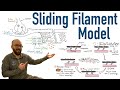
Understanding Muscle Contraction: The Sliding Filament Model Explained
Explore the sliding filament model of muscle contraction, from brain signals to muscle movement. Discover the intricate biochemical process involved.
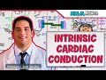
Comprehensive Guide to Cardiac Electrophysiology and Heart Conduction System
Explore the intrinsic electrical properties of the heart, including automaticity, nodal and contractile cells, and the detailed conduction pathway from the SA node through the Purkinje fibers. Understand how action potentials are generated and propagated, and how ion channels contribute to cardiac muscle contraction and relaxation.

Understanding the Three Muscle Types: Skeletal, Cardiac, and Smooth
Discover the differences and functions of skeletal, cardiac, and smooth muscle types in the human body.
Exploring the Three Types of Muscle: Skeletal, Cardiac, and Smooth
Dive into the differences between skeletal, cardiac, and smooth muscle and learn how they function in the body!
Understanding the Three Types of Muscle: Skeletal, Cardiac, and Smooth
Learn about the differences and similarities between skeletal, cardiac, and smooth muscles in this informative video with Dr. Mike.
Most Viewed Summaries

A Comprehensive Guide to Using Stable Diffusion Forge UI
Explore the Stable Diffusion Forge UI, customizable settings, models, and more to enhance your image generation experience.

Kolonyalismo at Imperyalismo: Ang Kasaysayan ng Pagsakop sa Pilipinas
Tuklasin ang kasaysayan ng kolonyalismo at imperyalismo sa Pilipinas sa pamamagitan ni Ferdinand Magellan.

Mastering Inpainting with Stable Diffusion: Fix Mistakes and Enhance Your Images
Learn to fix mistakes and enhance images with Stable Diffusion's inpainting features effectively.

Pamamaraan at Patakarang Kolonyal ng mga Espanyol sa Pilipinas
Tuklasin ang mga pamamaraan at patakaran ng mga Espanyol sa Pilipinas, at ang epekto nito sa mga Pilipino.

How to Install and Configure Forge: A New Stable Diffusion Web UI
Learn to install and configure the new Forge web UI for Stable Diffusion, with tips on models and settings.

If you found this summary useful, consider buying us a coffee. It would help us a lot!