Introduction
Muscle function is an essential aspect of our daily lives, yet we often overlook the complex biochemical processes behind muscle contraction. The sliding filament model explains how muscle fibers contract and relax, resulting in movement. This model involves a fascinating interplay between various chemical signals and structural components. In this article, we will break down the sliding filament model step by step, starting from the brain's action potential to the interaction between actin and myosin filaments.
Action Potential: The Beginning of Muscle Contraction
Muscle contraction initiates with signals from the brain. When a command is sent from the motor cortex, an action potential travels through neurons. Here’s a summary of the process:
- Brain Signal: The brain sends a signal down the spinal cord.
- Neurotransmitter Release: The signal travels to the axon terminal, leading to the release of neurotransmitters.
- Neuromuscular Junction: These neurotransmitters bind to receptors on the muscle fibers at the neuromuscular junction, directing the muscle to contract.
Excitation-Contraction Coupling
Before we dive deeper into the contraction mechanism, it's crucial to understand excitation-contraction coupling. This process involves the electrical signal triggering the calcium release necessary for muscle contraction. Here's the sequence:
- The action potential causes synaptic transmission, whereby sodium rushes into the postsynaptic muscle cell.
- This depolarization spreads along the muscle cell membrane (sarcolemma) and dives into the transverse tubules (T-tubules).
- The depolarization triggers the sarcoplasmic reticulum to release calcium ions into the muscle fibers.
The Sliding Filament Model
The heart of muscle contraction lies within the myofibrils, microscopic structures within muscle fibers containing two critical protein filaments: actin (thin filament) and myosin (thick filament). The interaction between these filaments is what causes muscle contraction through a process often referred to as the sliding filament model. Let’s explore this model in detail.
Step 1: Binding Sites Exposure
In a resting muscle, binding sites on actin are obstructed by tropomyosin, preventing myosin from attaching. The presence of calcium ions is essential for contraction:
- When calcium ions bind to troponin, tropomyosin shifts, exposing binding sites on actin.
Step 2: Cross Bridge Formation
Once the binding sites are available:
- Myosin heads attach to the exposed sites on the actin filaments, forming what we call a cross bridge.
- This interaction is crucial for the contraction process as it sets the stage for the power stroke.
Step 3: Power Stroke
This is where the actual contraction occurs:
- The myosin heads pull the actin filaments toward the center of the sarcomere, which shortens the muscle.
- This action is energy-consuming, utilizing ATP (adenosine triphosphate) to power the pull.
Step 4: Release and Reset
After the power stroke, the muscle must reset for the next contraction:
- ATP binds to the myosin head, causing it to detach from actin, which ends the cross bridge.
- The energy from ATP is used to reset the myosin head back into its high-energy state to prepare for another contraction.
Step 5: Continuous Contraction
If calcium ions remain present:
- The cycle of forming cross bridges, power stroke, and reset continues.
- Muscles can sustain contractions as long as there are signals for calcium release and adequate ATP is available.
Muscle Relaxation
Muscle relaxation occurs when:
- Neural signals stop, leading to calcium ions being pumped back into the sarcoplasmic reticulum.
- Without calcium, tropomyosin returns to its active position, blocking the binding sites on actin, and the muscle relaxes.
Conclusion
The sliding filament model of muscle contraction highlights the intricate and dynamic process behind muscle movement. It starts with an action potential from the brain and follows a detailed sequence involving neurotransmitter release, excitation-contraction coupling, and the interaction between actin and myosin filaments. Understanding this model not only deepens our appreciation for muscular function but also underscores the biological complexity that allows us to perform everyday movements effectively.
when it comes to our muscles we don't spend a lot of time thinking about what happens at the
chemical level uh whenever we do movements but it turns out it's a somewhat complicated and i think
fascinating process we call it the sliding filament model because the filaments are pulling or
sliding across each other of course every time we move our muscles it starts in the brain and the signal
down to the muscles to get them to contract we're going to start with an action
potential from the brain and work our way down to the sliding filament model where the actual proteins are pulling on
each other to contract the muscle let's jump to the white board and get started all right so before we get into the
sliding filament steps themselves we have to take a look at something called excitation contraction coupling which is
this idea that for a muscle to contract we first need a signal or an excitation
that's going to come from the brain the brain is going to send a signal down through the spinal cord
out through a nerve to whatever muscle it is that we're trying to move and then the muscle can contract as a
response to that so in our diagram here we have those two cells we have a neuron and this is the axon terminal of a
neuron we have a synapse the connection between this neuron and this muscle cell now
because it's a neuron and a muscle cell sometimes we also call this a neuromuscular junction but it's just a
synapse and that axon terminal of course has some vesicles with neurotransmitters
there's some receptors on the postsynaptic cell which is our muscle fiber
and our muscle fiber is going to have several organelles that we're going to label here
this first one the transverse tubule that goes down into the muscle cell as well as the sarcoplasmic reticulum
which is going to store calcium to be released as part of this process now i didn't draw all of the
sarcoplasmic reticulum i sort of cut it off here so i have room to draw and write other things in the diagram
and underneath the sarcoplasmic reticulum are the myofibrils which contain
the myofilaments so these are the myofilaments here and we have two of those of course the actin and the myosin
and that's kind of our end goal is to get them to pull on each other and shorten the length of the sarcomere
which is the space between this z line and this other z line and as that happens
our muscles contract that's our end goal here but it all starts with the brain sending action potentials through
neurons which is where we're going to start right now so first we have this action potential
it's come down the axon now it's made it to the axon terminal that's going to cause the vesicles to release the
neurotransmitters which are going to bind with the receptors on the postsynaptic side down here
that's going to cause sodium to rush into the postsynaptic cell through a process called synaptic
transmission and if you need to check out my videos on action potentials and on synapses which
i'll link to the description down below now once this muscle cell depolarizes right here because of the sodium rushing
in that's going to cause more sodium channels to keep opening down the length
of the neuron which causes that new action potential to travel down the sarcolemma the cell
membrane of the muscle eventually that signal is going to make it to a t tubule now i just have one t
or transverse tubule drawn in here but there's really lots of them throughout the muscle cell and they're all going to
be conducting the signal down into the muscle cell so the signal travels down into the transverse tubule
down to the sarcoplasmic reticulum which like i said is going to be filled with calcium ions that as soon as the signal
gets there these calcium ions are going to get released out into
the myofibrils and that all started with the action potential that through this chain of events now has caused the
sarcoplasmic reticulum to leak calcium ions into the myofibrils to interact with the myofilaments
and in the presence of calcium the myofilaments will start grabbing onto each other
and contracting and then we get something like this see how they're much closer together
the length of the actin and the length of the myosin didn't change but as they pulled on each other the
length between the z lines from there to there has decreased and now we have a contracted
muscle and as long as there's signals being sent from a neuron and therefore calcium being released
from the sarcoplasmic reticulum we're going to have contracted muscles so just a quick recap
of all that so far it all starts with the brain the motor cortex in the brain sending a signal
down through the spinal cord out through a nerve and eventually that's going to make it to the end of a
neuron that connects to a muscle cell so we start with an action potential that's going to cause synaptic
transmission in the neuromuscular junction that signal is going to travel down
along the sarcolemma down into the transverse tubules where it's going to interact with the sarcoplasmic reticulum
causing it to release calcium ions in the presence of calcium ions the actinomycin
will contract like that relax contract relax contract relax contract sorry we don't
have a huge animation budget we just do two frame animations here okay now let's take a deeper dive
into the sliding filament part of this which is where the filaments grab onto each other and how is that all regulated
by calcium so let's zoom in to this section right in here
all right so here we have our two filaments we have myosin in pink that's our thick filament and we have
actin in purple here that's our thin filament if you look closely you'll see green dots you see green dots on the
myosin heads as well as green dots on all these circles which are the actin molecules
those are binding sites this is where the myosin head can latch onto or bind with the actin
molecule throughout this video i'm going to use the non-scientific term of grab like the
myosin head grabs onto the actin but a better term to use really here is bind it's going to chemically bind
with the actin molecule whenever it pulls on it but we have a problem the binding sites are actually covered
up by a molecule called tropomyosin you'll see it here i just drew it in brown and notice how it's hard to find
the green dots on there you have to look really closely now that's because this molecule that i drew in brown
here this tropomyosin has roped off or blocked off the binding sites and i think about that
like it's roped off like if you go somewhere and the seats are roped off reserved for somebody else tropomyosin
is roping off and has rope in there right trope ropomyosin it's blocking the binding
sites meaning our muscle can't contract right now we have one more little molecule on there
and i do that in yellow and that's called troponin troponin's kind of connected to the tropomyosin and they're
going to be interacting with each other in a second so again we have myosin
we have the myosin heads we have the actin molecules in purple the green binding sites which is where
the myosin heads can grab onto the actin those of course are blocked by the tropomyosin that's that kind of brown
looking rope right there and we have troponin which i have here in yellow and we'll
see what that does in a second so at this stage we're sort of in the off setting right the muscle's not
contracted yet we're about to contract the muscle and i'm going to call that stage one here in
just a moment okay now let's assume that the sarcoplasmic reticulum has released calcium ions right we've
had a neuron send an action potential from the brain down to this muscle cell the
sarcoplasmic reticulum releases its calcium and here's what's going to happen some
of that calcium is going to bind with the troponin so these orange calcium ions are going to bond with the yellow
troponin right there and that's going to cause the tropomyosin to actually peel
back out of the way and if you look closely at our diagram now you'll see there's still tropomyosin
there but notice how it's not blocking the binding sites you can see those little green dots on
there pretty clearly now because the tropomyosin has been pulled back and that happened whenever the
calcium ions binded with the troponin which pulls back the tropomyosin
so the binding sites are exposed and now the myosin heads are going to be able to form a connection to them this is sort
of the on state now our muscle's about to contract but that's going to take a few stages
for it to happen notice in our diagram now that the myosin heads are physically connected
to the actin molecules their green binding sites are lined up with each other
right there and so this is latched on we call this forming a cross bridge i think of this as the grab stage the
myosin grabs onto the actin really it binds with it and we say that the myosin head forms a
cross bridge a bridge connects two things together and so we call this sort of a bridge a
cross bridge the myosin head has formed a cross bridge between the myosin molecule and the
actin filament once we've formed a cross bridge that myosin head now is going to pull
and we call the pole this is the actual scientific name for it we call the pull the power stroke so
turn on grab pull i think these stages are a lot easier to remember if you think of them
like that turn on grab pull we'll see what happens next all right we've pulled now and we
need to reset so that we can pull again or just to relax the muscle if we're not going to contract it anymore
so this stage has two parts to it one is to release and the second is to reset atp is used
in this process so contracting our muscles is pretty atp intensive
our brain along with our muscles use more energy than any part of the body and this is one reason why
every time we have a grab and pull and release cycle here we have to use up an atp
molecule you need that energy from it for this to happen so the atp does two things it's
going to break the cross bridge and then it uses up its energy to reset the myosin head back to where it was
i like to think of this like a mouse trap the mouse trap you have to set the mouse trap right you
have to put energy in to pull the mouse trap back but once it's set
it doesn't take any more energy from you it just takes this is kind of morbid i guess
it just takes the mouse to crawl up onto it and then it snaps but the energy's already there you
already give it the energy whenever you back the bar or the myosin head here it has the
energy now it's just ready to release it same thing here with this
the atp is what's going to be used to one break the cross bridge and then two to reset or pull back to
the high energy state the myosin head and i think a mousetrap is a good metaphor for it because
this is where the energy from the atp is consumed not the power stroke which is where you would think like
oh we use the energy we really use the energy to reset the myosin head so a little bit later it can
do the power stroke so we've reset one of two things can happen here if there's calcium still
present it's just gonna keep doing this over and over and over again repeating steps two three and four that myosin
head is gonna grab onto the actin it's going to pull and then it will release and reset in the presence of atp
grab pull release and reset grab pull release and reset unless there's no more calcium
then we're done as long as there's calcium present in other words as long as the brain keeps sending
signals to the sr to keep releasing its calcium this muscle cell is going to keep
contracting and contracting and contracting as long as it can this would be like sustaining a muscle contraction
over a long period of time and you're not like letting go but we don't want all of our muscles to stay
contracted all the time that would be really bad so we have to be able to relax muscles as well and what happens
there well calcium is just going to get re-pumped back into the sarcoplasmic reticulum so
that it can't interact with this process anymore and that's going to turn off the muscle cell
because remember if there's no calcium present the tropomyosin will block off the
binding sites and the muscle can't contract anymore the muscle then will relax
and it'll be ready to contract again whenever we have more calcium released from the sr but until then
we're back to our relaxed state ah so to recap all of that we've got our myosin and our actin filaments
when the presence of calcium that's going to turn this on basically the sr releases the calcium it bonds with the
troponin the troponin pulls back the tropomyosin revealing the binding sites
the myosin heads are going to grab on to the binding sites we call that forming a cross bridge
they're going to pull on the actin molecule we call it the power stroke and then the myosin head and the
presence of atp is going to release and reset back to its excited state that process
of grabbing pulling releasing resetting grabbing pulling releasing resetting that's going to keep happening
as long as there is calcium present from the sr whenever we're ready to relax the muscle
the sr is going to pump the calcium back into the sarcoplasmic reticulum and once that calcium is gone
then the tropomyosin covers up the binding sites and we're back to our relaxed
state and of course this whole process like i said has to start with an action potential
synaptic transmission conduction down the sarcolemma into the t tubule causing the sr to release calcium
and then it goes through all of that stuff that we just did to have a nice contracted muscle now if
you want to test your understanding take a moment and pause the video describe what happens in each of the five steps
of the sliding filament model without any of the text on here as a guide if you can explain what
happens in each of those five steps then you have a pretty good understanding of the sliding filament model
of muscle contraction now here's that text back again if you want to check and see how you did
all right can you find the a in filament where's the a can you point to the a yeah good job
good job yeah what's over that can you point can you find can you find an m can you
find an m good job have we even learned that one yet how do you know where an m is
Heads up!
This summary and transcript were automatically generated using AI with the Free YouTube Transcript Summary Tool by LunaNotes.
Generate a summary for freeRelated Summaries
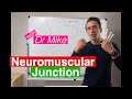
Understanding the Neuromuscular Junction: Mechanics of Muscle Contraction
Explore the neuromuscular junction process, including neurotransmitter action and muscle contraction mechanics.
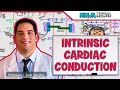
Comprehensive Guide to Cardiac Electrophysiology and Heart Conduction System
Explore the intrinsic electrical properties of the heart, including automaticity, nodal and contractile cells, and the detailed conduction pathway from the SA node through the Purkinje fibers. Understand how action potentials are generated and propagated, and how ion channels contribute to cardiac muscle contraction and relaxation.
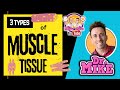
Exploring the Three Types of Muscle: Skeletal, Cardiac, and Smooth
Dive into the differences between skeletal, cardiac, and smooth muscle and learn how they function in the body!
Understanding the Three Types of Muscle: Skeletal, Cardiac, and Smooth
Learn about the differences and similarities between skeletal, cardiac, and smooth muscles in this informative video with Dr. Mike.
Understanding the Three Muscle Types: Skeletal, Cardiac, and Smooth
Discover the differences and functions of skeletal, cardiac, and smooth muscle types in the human body.
Most Viewed Summaries

A Comprehensive Guide to Using Stable Diffusion Forge UI
Explore the Stable Diffusion Forge UI, customizable settings, models, and more to enhance your image generation experience.

Kolonyalismo at Imperyalismo: Ang Kasaysayan ng Pagsakop sa Pilipinas
Tuklasin ang kasaysayan ng kolonyalismo at imperyalismo sa Pilipinas sa pamamagitan ni Ferdinand Magellan.

Mastering Inpainting with Stable Diffusion: Fix Mistakes and Enhance Your Images
Learn to fix mistakes and enhance images with Stable Diffusion's inpainting features effectively.

Pamamaraan at Patakarang Kolonyal ng mga Espanyol sa Pilipinas
Tuklasin ang mga pamamaraan at patakaran ng mga Espanyol sa Pilipinas, at ang epekto nito sa mga Pilipino.

How to Install and Configure Forge: A New Stable Diffusion Web UI
Learn to install and configure the new Forge web UI for Stable Diffusion, with tips on models and settings.

If you found this summary useful, consider buying us a coffee. It would help us a lot!