Overview of Atomic Force Microscopy (AFM)
- Definition: AFM is a type of scanning probe microscopy that provides high-resolution imaging of surfaces at the nanoscale.
- Functionality: It works by scanning a sharp tip over a sample surface, measuring the forces between the tip and the sample to create detailed 3D images.
Key Components of AFM
- Cantilever: A flexible beam that holds the tip and bends in response to forces from the sample.
- Laser and Photodetector: Used to measure the deflection of the cantilever, which is crucial for imaging.
How AFM Works
- Sample Interaction: The tip interacts with the sample surface, and the resulting forces cause the cantilever to deflect.
- Data Collection: The deflection is measured using a laser beam reflected off the cantilever into a photodetector.
- Image Construction: The data collected is processed to create a 3D image of the sample surface.
Advantages of AFM
- High Resolution: Capable of imaging at the atomic level.
- Versatility: Can be used for a variety of materials and biological samples.
- Non-destructive: Does not damage the sample during imaging.
Applications of AFM
- Material Science: Analyzing surface properties of materials, which is also a key aspect of Understanding Microorganisms: Types of Microscopes and Their Applications.
- Biology: Studying biological samples at the nanoscale, similar to techniques discussed in Understanding Quantum Mechanics: A Comprehensive Guide.
- Nanotechnology: Fabricating and characterizing nanostructures, a topic that overlaps with Understanding Electromagnetism: The Basics of Forces, Mass, and Charge.
Conclusion
- AFM is a powerful tool in modern microscopy, offering unique insights into the structure and properties of materials at the nanoscale. For more information and tutorials, subscribe to the channel.
MP3 कि एटॉमिक पोस्ट माइक्रोस्कोप कॉल एग्जाम सिस्टमिक रिफॉर्म्स माइक्रोस्कोप इजहार
रिलेशंस कैन इंप्रूव माइक्रोस्कोप थ्री इडियट टूल फॉर इमेजिंग मेजरिंग नुस्खे चैनल सब्सक्राइब टो
हाउ टो मेक पोस्ट माइक्रोस्कोप वर्क्स इन थे आइडिया आफ प्रोड्यूजिंग मिर्च वाइफ फिजिकली पुशिंग हेर कैन डिलीवर टिप्स
अगेंस्ट द सैंपल है जो व्यक्ति मूवमेंट आफ डिफरेंट इलनेसेस एंड कन्वर्टेड इनटू थ्री डाइमेंशनल इमेज
ऑफ द सैंपल सर्विस है कि एटॉमिक मैस माइक्रोसॉफ्ट कंसिस्ट्स आफ अप माइक्रो स्किल कैंटिलीवर ऑफ लंदन और
उन्हें ज़ूम ज़ूम विद अश आफ बन्रट इसको लुट रेडियो सॉफ्ट टॉयज फ्रॉम दिस इज पेज ओनली फॉर रेजिडेंट्स ले ले ले फोटो ले
हैं The Light कलेक्टेड बाय थिस फोटो एडिटर्स एंड लाइव्स विद द हेल्प ऑफ कंप्यूटर डिवाइस एंड 3D इमेज सैंपल सरफेस
कैन बे कंस्ट्रक्टेड ओं में वृद्धि कैंटिलीवर टिप्स ब्रोट इनटू थे प्रॉक्सिमिटी आफ सैंपल सरफेस द फ्यू सचिव
व से इलेक्ट्रोस्टेटिक फोर्स मांगी डिपो से टैब्स विच अराइज ड्यू टो फिजिकल इंटरेक्शन बिटवीन subscribe to subscribe
the Channel द चेंजिंग एंड अकॉर्डिंग्ली ए कि वे कैन फाइंड द अमाउंट ऑफ डिफलेक्शन प्रोड्यूस इन दिशाओं रिलेटिड लेजर बीम बाय
एनालाइजिंग विच फोटो डिटेक्टर्स कलेक्टिंग एक पर्टिकुलर Play Services कैन बे डन विद थे हेल्प आफ कंप्यूटर कंट्रोल डिवाइस
मैं अनिल 120 डिफलेक्शन विल हेल्प उस इंप्रूव सिंगर 3D इमेज ऑफ द सैंपल सर्विस सर्विस एनिमेशन विल गिव एनी आइडिया अबाउट
हाउ टो मेक ए माइक्रोस्कोप विल वर्क ओं थे सॉफ्टवेयर भी है वह समाधान तेजस एंड डिसएडवांटेजेस आफ एटॉमिक पोर्स
माइक्रोस्कोप फिसड्डी एडवांटेजेस इन टू-डू सलूशन ऑफ बट व्हेन वॉर्न इन कंडीशन कैन बे अचीव्ड थ्री डाइमेंशनल जॉब स्किन कैन बे
यूज्ड फॉर यू ऑल सबस्क्राइब और मॉनिटर सब्सक्राइब करें सब्सक्राइब डिपोजिट पॉइंट सब्सक्राइब वेरी वेरी सिंपल सब्सक्राइब टो
एटॉमिक पोर्स माइक्रोस्कोप थैंक यू फॉर वाचिंग माय वीडियो को लाइक दिस वीडियो दिन Please subscribe The Channel टाइमली
इंटरवेंशन आफ न्यू वीडियो अपलोड टोटके चैनल थैंक यू थैंक यू वेरी मच
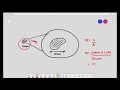
Atomic Force Microscopy (AFM) is a type of scanning probe microscopy that provides high-resolution imaging of surfaces at the nanoscale. It works by using a cantilever with a sharp tip that physically interacts with the sample surface. As the tip moves across the surface, it experiences forces that cause it to deflect, which is measured to create a three-dimensional image of the sample.
The main components of an Atomic Force Microscope include a cantilever with a sharp tip, a laser beam to detect cantilever deflection, a photodetector to measure the laser's position, and a computer to process the data and generate images. These components work together to provide detailed information about the surface topography and properties of the sample.
Atomic Force Microscopy offers several advantages, including the ability to image samples in air or liquid environments, high-resolution imaging down to the atomic level, and the capability to measure various properties such as mechanical, electrical, and magnetic characteristics of materials.
Despite its advantages, Atomic Force Microscopy has limitations, such as slower imaging speeds compared to other microscopy techniques, potential damage to soft samples due to the tip's interaction, and the requirement for careful calibration and operation to obtain accurate results.
AFM is widely used in various fields, including materials science, biology, and nanotechnology. It can be employed to study surface roughness, measure mechanical properties of materials, analyze biological samples at the nanoscale, and develop new nanostructured materials.
To create a 3D image using AFM, the cantilever tip scans the sample surface while measuring the deflection caused by interactions with the surface. This data is collected and processed by a computer to construct a detailed three-dimensional representation of the sample's topography.
To learn more about Atomic Force Microscopy, you can watch educational videos, read scientific literature, and explore online resources that provide tutorials and detailed explanations of AFM principles, applications, and techniques.
Heads up!
This summary and transcript were automatically generated using AI with the Free YouTube Transcript Summary Tool by LunaNotes.
Generate a summary for freeRelated Summaries
Understanding Microorganisms: Types of Microscopes and Their Applications
Explore the fascinating world of microorganisms and the various microscopes used to observe them in detail.
Understanding Cell Structure: Basics of Microscopy and Magnification
This video explains the fundamentals of cell microscopy, focusing on how microscopes magnify specimens to reveal cell structures. Learn about light microscopes, the concept of magnification, unit conversions, and how light interaction enables us to see microscopic objects.

Comprehensive AQA Atomic Structure Revision Guide Explained
This video provides a detailed overview of AQA atomic structure, covering key concepts such as atomic models, ions, isotopes, electron configurations, ionization energy, and mass spectrometry. Ideal for students preparing for AQA chemistry exams, it explains complex topics with clear examples and practical insights.

Understanding Atomic Structure: From Atoms to Subatomic Particles
Explore the fascinating journey of atomic theory from ancient Greek philosophy to modern discoveries of subatomic particles. Learn how atoms are structured, the role of protons, neutrons, and electrons, and the significance of isotopes and ions in chemistry.

Understanding Atoms: Structure, Particles, and Elements
This video explores the fundamental building blocks of matter, known as atoms, detailing their structure, components, and the concept of ions. It explains the roles of protons, neutrons, and electrons, as well as how elements are represented in the periodic table.
Most Viewed Summaries

A Comprehensive Guide to Using Stable Diffusion Forge UI
Explore the Stable Diffusion Forge UI, customizable settings, models, and more to enhance your image generation experience.

Kolonyalismo at Imperyalismo: Ang Kasaysayan ng Pagsakop sa Pilipinas
Tuklasin ang kasaysayan ng kolonyalismo at imperyalismo sa Pilipinas sa pamamagitan ni Ferdinand Magellan.

Mastering Inpainting with Stable Diffusion: Fix Mistakes and Enhance Your Images
Learn to fix mistakes and enhance images with Stable Diffusion's inpainting features effectively.

Pamamaraan at Patakarang Kolonyal ng mga Espanyol sa Pilipinas
Tuklasin ang mga pamamaraan at patakaran ng mga Espanyol sa Pilipinas, at ang epekto nito sa mga Pilipino.

How to Install and Configure Forge: A New Stable Diffusion Web UI
Learn to install and configure the new Forge web UI for Stable Diffusion, with tips on models and settings.

If you found this summary useful, consider buying us a coffee. It would help us a lot!