Wat zijn eiwitten?
Eiwitten vormen de bouwstenen van ons lichaam en zijn opgebouwd uit aminozuren. Na het menselijk genoomproject richt de wetenschap zich nu op het proteoomproject, dat onderzoekt hoe eiwitten zijn samengesteld en hoe ze er driedimensionaal uitzien. Voor een dieper begrip van de basiscomponenten van eiwitten, kun je meer lezen over Understanding the Structure of DNA: Key Components and Functions.
Aminozuren: de bouwstenen van eiwitten
- Eiwitten bestaan uit 20 verschillende aminozuren.
- Elk aminozuur heeft een centrale alfa-koolstof met een amino groep (links), een carboxyl groep (rechts) en een unieke R-groep die de eigenschappen bepaalt.
- Aminozuren komen uit ons dieet; ons lichaam breekt eiwitten af tot aminozuren en bouwt ze weer op tot nieuwe eiwitten. Voor meer informatie over de verschillende soorten biomoleculen, kun je onze gids over Understanding Biomolecules: A Comprehensive Guide bekijken.
Hoe worden eiwitten gevormd?
- Aminozuren verbinden zich via een proces genaamd dehydratie synthese, waarbij water wordt afgesplitst en covalente bindingen ontstaan.
- Deze ketens van aminozuren heten polypeptiden.
- Ribosomen in cellen zorgen voor het samenvoegen van aminozuren tot polypeptiden. Voor een beter begrip van de rol van cellen in dit proces, lees Understanding the Structure and Function of the Cell: A Comprehensive Overview.
Chemische eigenschappen van aminozuren
- Aminozuren kunnen polair (wateraantrekkend) of apolair (waterafstotend) zijn.
- Positief en negatief geladen aminozuren trekken elkaar aan, wat invloed heeft op de eiwitvouw.
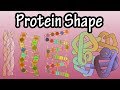
De vier structuurniveaus van eiwitten
- Primaire structuur: de lineaire volgorde van aminozuren.
- Secundaire structuur: alfa-helixen en beta-sheets, gestabiliseerd door waterstofbruggen.
- Tertiaire structuur: driedimensionale vouwing door interacties tussen R-groepen, inclusief hydrofobe en hydrofiele gebieden en zwavelbruggen.
- Quaternaire structuur: samenvoeging van meerdere polypeptideketens tot een functioneel eiwit, zoals hemoglobine. Voor meer inzicht in de eiwitstructuur en de rol van RNA in dit proces, zie The Essential Roles of RNA in Genetics and Protein Synthesis.
Het belang van eiwitvouwing
- De specifieke driedimensionale vorm bepaalt de functie van het eiwit.
- Veranderingen in temperatuur of pH kunnen eiwitten denatureren, waardoor ze hun functie verliezen.
Zelf eiwitten vouwen met Foldit
- Foldit is een interactief spel waarin je polypeptiden kunt vouwen om stabiele eiwitstructuren te creëren.
- Spelers leren de regels van eiwitvouwing en dragen bij aan wetenschappelijk onderzoek.
- Foldit-gebruikers hebben al geholpen bij het ontrafelen van belangrijke enzymstructuren, zoals die van HIV.
Conclusie
Eiwitten zijn essentieel voor het leven en hun complexe structuren bepalen hun functies. Met tools zoals Foldit kunnen ook niet-wetenschappers bijdragen aan baanbrekend onderzoek door eiwitten te vouwen en zo nieuwe inzichten te bieden in de biochemie.
Hi. It's Mr. Andersen and in this video I'm going to talk about proteins. Proteins are incredibly important because that's what we're made up of. When you look at me you're looking at my proteins. We just completed the human genome project and we
figured out the DNA in a typical human but now we're headed into what's called the proteom project where we try to figure out what these proteins made up of and what do they look like three dimensionally. This used to be an incredibly hard process. This is John Kendrew
here putting together a model for myoglobin and he had to do it by hand. We now do a lot of this with computers. But you can help. At the end of this video I'm going to show you a program called Foldit and you can actually build and fold proteins that are going to
be used in scientific research. And so it's a really cool thing. And it's a really cool time in reference to proteins. But let's start by building a little bit of knowledge. Proteins are made of amino acids. And most seventh graders understand this. They're the building
blocks of proteins. But where do we get those amino acids? We get them in our diet. And so basically we eat proteins. We break them down into amino acids and then we can weave those back together again into the proteins that make us. And so when you're looking at
me, the amino acids in my skin, used to be part of my food. And so I literally am what I eat. Now here's five different amino acids. There are a total of twenty that we use in life, but here's five basic amino acids. When I show this to my students they tend to get
a little bit overwhelmed because it's too much chemistry here on one page. But let's try to figure out the things that are the same on this page. And so basically if we were to look at the middle of each of these we find that there's a carbon with a hydrogen
attached to it. We call this carbon alpha carbon. It's going to sit right in the middle. What else is the same in every amino acid? Well on the left side we have a nitrogen attached to two hydrogens. We call this an amino group. And then if we look on the right side we have
what's called a carboxyl group. And so basically every amino acid is going to have these three similar parts. And so the only thing that's going to be different is going to be what comes off the bottom. And we call that the R group. And so all amino acids are the same
except what comes off here. And that gives it different properties. And so let's kind of see how they're put together. And so when I build proteins inside my body, I'm doing that with amino acids. And we use something called dehydration synthesis to do that. And
so let me move this one over here. Basically what we do is we position one amino acid right next to an amino acid. You can see here that there is two hydrogens and an oxygen here and you know that in chemistry if we have two hydrogens, H2O that's simply a water.
And so what we can do is we can lose that water. Now it's not as simple as that. This whole thing sits inside a ribosome. So there's a giant enzyme around the outside of it. But let's attach another one. So now we bring another one right next to it. You can see
that the hydroxyl group or the carboxyl group is attaching to the hydrogen. We're going to lose a water and then we're going to form another covalent bond. And then we put another one next to it. We lose another water, we form a covalent bond and we do that again
and now we have what's called a polypeptide. And so each of these individually are called a peptide. But if we attach them all together we have a polypeptide. And you can see that the strand across the top looks the same. It looks uniform, but the only thing that's
going to be different in each of these is going to be the R group, the trails off the bottom. So where does this occur? Well basically these are the amino acids. This would be the ribosome in a cell and all life does this. Basically you have these little tRNAs that
will bring their amino acids in and then we attach them together. And when you have a bunch of amino acids attached together, you have a polypeptide. And that polypeptide will eventually fold into a protein. And so here's our five amino acid sequence right here. These
are each going to have different chemical properties. And so for example, this one right here, threonine is going to be polar. What that means is it's going to have a charge. If we look at alanine right down the way this is going to be nonpolar. And so why is that
important? Well if you're polar then you're hydrophilic. That means that water is going to be attracted to you. In other words we're going to find threonine in the presence of water. But alanine, since it's got this methyl group right here, it's not going to be attracted
to the water and so it's going to hide from the water. Or if we look at the aspartic acid, it's going to have a negative charge. And if we look at the lysine over here it's going to have a positive charge. And so these two things or these two amino acids are found
in the same polypeptide and they're going to try to get next to each other because the positive and the negative are going to attract. And so what you end up getting is a three dimensional protein. The middle part, so this brownish tan part in the middle is going to
be the back bone. And that's going to be again made up of all of the parts of the amino acid that are the same so the amino, the alpha and the carboxyl group over and over and over again, but all of these things on the outside that are trailing off are going to be the
R groups. These are going to be these residue groups that kind of fold off the end. And so this right here would be a polypeptide. This would be a number of different amino acids attached together and these usually have thousands of amino acids in a typical
protein like hemoglobin would be an example of one that's found in your blood. And so basically this will fold into a three dimensional shape. And what I tell students is a polypeptide is just going to be this sequence of those amino acids and once it's folded into a specific
shape then we can call it a protein. And so the first, maybe you have read it, there are four levels of structure in a protein. And so let me talk you through that first and then I've got a little model that will hopefully help. And so the primary structure is going
to be the order that those amino acids are bonded together. The next thing we have are what are called alpha helixes and beta pleated sheets. And we call that the secondary structure. And so a helix looks like this. A beta pleated sheet is going to be two sides that are attached
to each other and these little dots in the middle are going to be hydrogen bonding between adjacent sides of that polypeptide. And so this is the structure that comes out first. It's going to be linear. Next we have the alpha helixes or the beta pleated sheets.
And then we have the tertiary structure. The tertiary are the third level of structure, is going to be all of those R groups interacting and so maybe we have one that's hydrophobic. It'll hide to the middle or hydrophilic on the outside. We'll have disulfide bonds. We'll
have positive attracting to negative. This is the third level of structure. And then finally we have quaternary structure. Maybe we have this one polypeptide together or this protein together with another protein. So hemoglobin's an example of that made of a
number of different subunits. And so here's my little model. And so if you look at this model you can think of this being a polypeptide. So it's made up of amino acid after amino acid and then it's going to have all the R groups on the underside. Those are going to
be the only things that are different, all these R groups coming off the bottom. And so basically, primary structure like this. Secondary structure is going to be the alpha helixes and the beta pleated sheets. And so an alpha helix will look like this. What's
holding that in place is simply going to be the hydrogen bonds. And then we're going to have a beta pleated sheet. A beta pleated sheet might look like something like that. So there's going to be hydrogen bonds between here and here. But maybe this right here is
a real hydrophobic R group, and it's going to fold right to the middle and then this might be hydrophilic. It's going to fold to the outside. We might have positive attracted to negative then we eventually have a three dimensional shape of a protein. Now this may
combine with other proteins. But what's cool about proteins is their structure fits their function. If it doesn't have this structure, if we heat it up, if we cool it, if we change the acidity, basically it will fold apart. We call that denature and then it doesn't
work anymore. And so I said at the end you could help in science. So there's a program called Foldit. I'm going to launch it and I'll be back in just a second. And so in this program what you're given is a simple polypeptide. So we have two amino acids and then this is
going to be the R group. And what you do in this video game is you try to make the R groups happy. So I've already cleared level one. So let's go on to level 2. You can download this for Mac, Lynx and Windows. Let me quickly turn this one around. You can see here that
we have a couple of, let me get the help out of the way, you have a couple of different amino acids and then their R groups. I can pull those apart and they're going to be a little bit happier and then I can clear the level. And so what are you really doing. As
I play this game I can talk, basically what you're doing is you're learning the rules of protein folding, but these problems are going to get harder and harder and harder. You can see an alpha helix here. And so what you can do is you can get really good at folding
these proteins. And what's neat about this is people are playing Foldit hour and hour after hour. And it hit the news last year where a couple of protein folders, probably a team of protein folders decoded the shape of a really important enzyme in HIV infection.
And so it's plausible that in the future gamers are going to win a Nobel prize for the work they do on protein folding. Because we know the primary structure of proteins, but we don't have any idea of how the three dimensional shape is put together. And computers are good
at this but it turns out that humans are maybe a little bit better. And so those are proteins and I hope that was helpful.
Eiwitten zijn de bouwstenen van ons lichaam, opgebouwd uit aminozuren. Ze spelen een cruciale rol in vrijwel alle biologische processen, waaronder enzymatische reacties, celstructuur en transport van moleculen. Hun specifieke functies zijn afhankelijk van hun complexe driedimensionale structuren.
Eiwitten worden gevormd door aminozuren die zich verbinden via dehydratie synthese, waarbij water wordt afgesplitst. Dit proces vindt plaats in ribosomen, waar aminozuren worden samengevoegd tot polypeptiden, de voorlopers van eiwitten.
De vier structuurniveaus van eiwitten zijn: 1) Primaire structuur - de lineaire volgorde van aminozuren; 2) Secundaire structuur - vormen zoals alfa-helixen en beta-sheets; 3) Tertiaire structuur - de driedimensionale vouwing door interacties tussen R-groepen; 4) Quaternaire structuur - de samenvoeging van meerdere polypeptideketens tot een functioneel eiwit.
De specifieke driedimensionale vorm van een eiwit bepaalt zijn functie. Als eiwitten denatureren door veranderingen in temperatuur of pH, verliezen ze hun functie, wat kan leiden tot ernstige gevolgen voor de cel en het organisme.
Foldit is een interactief spel waarin spelers polypeptiden kunnen vouwen om stabiele eiwitstructuren te creëren. Door het spel leren spelers de regels van eiwitvouwing en dragen ze bij aan wetenschappelijk onderzoek, zoals het ontrafelen van enzymstructuren, waaronder die van HIV.
De chemische eigenschappen van aminozuren, zoals polariteit en lading, zijn essentieel voor hun rol in eiwitvouwing. Voor meer informatie kun je onze gids over biomoleculen bekijken, die dieper ingaat op de verschillende soorten en functies van biomoleculen.
Denaturatie van eiwitten, veroorzaakt door factoren zoals temperatuur of pH-veranderingen, leidt tot verlies van de specifieke structuur en functie van het eiwit. Dit kan ernstige gevolgen hebben voor cellulaire processen en de algehele gezondheid van een organisme.
Heads up!
This summary and transcript were automatically generated using AI with the Free YouTube Transcript Summary Tool by LunaNotes.
Generate a summary for freeRelated Summaries
Understanding Protein Structure: Primary to Quaternary Levels Explained
Explore the four levels of protein structure—primary, secondary, tertiary, and quaternary—and learn how each shape influences protein function. Discover how proteins fold, bond, and how denaturation affects their activity.
Comprehensive Guide to Recombinant Protein Expression and Structural Biology
Explore the essential techniques scientists use to express, purify, and analyze proteins. This guide covers recombinant protein expression, chromatography purification methods, and structural biology tools like X-ray crystallography and cryo-EM to connect protein form with function.
Understanding the Structure of DNA: Key Components and Functions
Dive into the fascinating structure of DNA, exploring its components and their vital roles in genetics.

Comprehensive Insights into EBI and Essential Bioinformatics Tools
Explore the pivotal role of the European Bioinformatics Institute (EBI) in managing diverse biological databases and discover key bioinformatics tools for sequence analysis, pattern recognition, and structural comparison. Understand the synergy between wet labs and dry labs in modern bioinformatics and how EBI supports genomic and proteomic research.

Comprehensive Guide to Molecular File Formats for Protein 3D Modeling
Explore the essential molecular file formats like PDB, mmCIF, CHARMM, MDL, and Mopac used in protein 3D structure modeling. Understand their specific sections, applications in crystallography and molecular dynamics, and learn about key file conversion tools to integrate diverse data sources effectively.
Most Viewed Summaries

A Comprehensive Guide to Using Stable Diffusion Forge UI
Explore the Stable Diffusion Forge UI, customizable settings, models, and more to enhance your image generation experience.

Kolonyalismo at Imperyalismo: Ang Kasaysayan ng Pagsakop sa Pilipinas
Tuklasin ang kasaysayan ng kolonyalismo at imperyalismo sa Pilipinas sa pamamagitan ni Ferdinand Magellan.

Mastering Inpainting with Stable Diffusion: Fix Mistakes and Enhance Your Images
Learn to fix mistakes and enhance images with Stable Diffusion's inpainting features effectively.

Pamamaraan at Patakarang Kolonyal ng mga Espanyol sa Pilipinas
Tuklasin ang mga pamamaraan at patakaran ng mga Espanyol sa Pilipinas, at ang epekto nito sa mga Pilipino.

How to Install and Configure Forge: A New Stable Diffusion Web UI
Learn to install and configure the new Forge web UI for Stable Diffusion, with tips on models and settings.

If you found this summary useful, consider buying us a coffee. It would help us a lot!