Comprehensive Overview of Skull Anatomy and Related Structures
Introduction
The video covers essential concepts related to the anatomy of the skull, including skeletal landmarks, cranial fossa, and the development of various structures. It provides a detailed examination of bones, sutures, and important anatomical landmarks, as well as the development of the tongue and associated muscles.
Key Topics Discussed
-
Skeletal Landmarks of the Skull
- Identification of major bones: frontal, parietal, temporal, occipital, sphenoid, and mandible. For a deeper understanding of the skeletal structure, refer to Understanding the Human Skeleton: A Detailed Overview of Bones and Structure.
- Important sutures: sagittal, lambdoid, and coronal sutures.
- Anatomical landmarks: terion, lambda, bregma, vertex, glabella, asterion, and inion.
-
Cranial Fossa
- Overview of the anterior, middle, and posterior cranial fossa. This section ties into the broader context of skull anatomy discussed in Understanding the Human Skeleton: Structure, Function, and Significance.
- Discussion of the structures contained within each fossa.
-
Development of the Tongue
- Explanation of the embryological development of the tongue from pharyngeal arches.
- Identification of intrinsic and extrinsic muscles of the tongue.
-
Muscles of Facial Expression
- Overview of muscles responsible for facial expressions, categorized by their location (around the eye, nose, and mouth).
- Innervation of these muscles by the facial nerve (cranial nerve VII).
-
Cervical Plexus and Ganglia
- Description of the cervical plexus and its branches, including cutaneous and motor branches. For more on the nervous system's role in anatomy, see Understanding the Brain: The Link Between Neuroanatomy and Personality.
- Discussion of the submandibular and otic ganglia, their functions, and connections to cranial nerves.
Conclusion
The video serves as a comprehensive guide to understanding the complex anatomy of the skull and its associated structures, providing valuable insights for students and professionals in the field of anatomy and medicine.
[Music] hello guys uh so now we shall be discussing all the important Concepts
that are related with the head okay so all the important topics in the head so first important topic which we shall be
discussing over here will be the landmarks of your skull okay will be the skeletal landmarks of your skulls
skeletal landmarks of the skull skeletal landmarks of the skull coming to the
skeletal landmarks of the skull first of all in this particular picture which you can see what are the different kinds of
Bones which you can see over here first of all we'll discuss that and then we shall discuss other different landmarks
over here first of all this particular bone which you can see over here is named as your frontal bone all of you
know this this is your frontal bone this bone over here is called as your paral bone paral bone is best visualized from
the top okay next this particular bone over here is called as your temporal bone and you see a bone on the back side
over here this is called as oxital bone oxital bone so how many bones you have got over here you have got the
frontal bone you have got the perial bone you have got the temporal bone and the occipital bone and in the same way
uh you see a bone over here right this particular bone right so this particular bone over here is called as phid
bone okay this bone over here is called as a sphenoid bone so you can also appreciate one very important thing that
the frontal parital temporal and sphenoid Bone all of them are meeting at this point you see so the point where
all the frontal peral temporal occipital and the sphenoid bone the point where all of them are meeting over here right
so this particular point over here is called as terion what is this called as this is called as a terion now if there
is a fracture of teron actually there is a middle menal veins vessels all of them are present behind this okay I will tell
what are the contents of the teron later on right so but you have to remember that this teron is a combination of all
these uh bones frontal peral temporal sphenoid temporal parital and frontal all these bones together form the terion
that is another important question next important anatomical landmark after terion right so it is
terion terion next important anatomical landmark after teron is if you if you look at this right so you know that this
is peral bone this is another peral bone this is your occipital bone the suture which you can see between the the two
peral bones over here this is called as what this is called as your sagital suture sagital suture in the same way
the suture that is present between the occipital bone and the paral bone this suture over here is called as lamboid
suture okay lamboid suture so the where is the place where the seital suture and lamboid suure both of them are joining
that is the area number four over here you see this is the place where both the seital switcher right and two lamboid
switches they're joining here right so this particular area over here is called as
Lambda so what is the area called as Lambda Lambda is nothing but the place where or the junction of lamboid and the
sagital switcher fusing at that region is called as Lambda now if you look at this right so this is your anterior part
this this is your posterior part right you know you know that what is this particular suture over here this
particular suture over here is called as seital suture and this particular suture which
I'm drawing right now with the red is called as coronal suture coronal
suture and what is this sagital suture so the place where sagital suture and coronal suture are fusing exactly at
this Point okay so this point over here is called as bregma what is this called this region is called as
bregma okay so what is bregma bregma is a place where both the sagital and the coronal sutes are fusing that is called
as brigma so so far you have learned what so far you have learned teron what is Lambda and you have learned what is
brigma now look at this what is vertex the next important anatomical landmark is vertex the
highest point of the neurocranium so this entirely is your neurocranium the topmost part the topmost part is exactly
here you see this is the point okay this is the topmost point topmost elevated Point okay so this topmost point over
here is called as vertex so the Superior part Superior part of neurocranium okay that is called vertex
for example if I'm putting all my fingers in this way right if I ask you what is the
topmost area this can be topmost this can be topmost so this is a topmost in this this finger the middle finger over
here is having the topmost point this is called as a Vertex okay so that is a vertex there which we were discussing
right now okay right next important anatomical landmark is that see you you even have you even have got a a swelling
in the frontal region yes right you see an anterior projection of the forehead so this particular anterior projecting
part of the forehead is called as glab this is called glabella so what is your glabella glabella is nothing but
the anterior projecting anterior projecting part of forehead part
of forehead okay anterior projecting part of forehead is called as glaba next important thing is that where is
terion here see this is your teron all of you know that right this is your teron okay now I'm telling you that this
is your frontal bone sphenoid bone temporal bone and what is this parital bone and down here occipital bone now
you see a point you see a point over here this is a point where this is a point where your uh peral bone your
temporal bone and oxital Bone all of them are fusing this point where temporal peral temporal
peral and oxital Bone all of them are fusing this point over here is called as asterion okay
asterion this point over here is called as asterion okay now after this asterion the next important point this is from
behind Okay this is from the posterior side so on the posterior side you see a point over here near the external
occipital protuberance so this Prominent Point of external oxital protuberance is called as Inon okay it is called as Inon
what is Inon Inon is nothing but your prominent part Prominent Point Prominent Point of
external oxital protuberance oxital protuberance okay Prominent Point of external oxital protuberance is
called as in on okay next important thing here uh you see this is the nose this is the nose over here right and I
can tell you that this is the root of the nose the root of the nose is called as Nason what is Nason Nason is nothing
but the root of the nose just above that you have that swelling called as glaba okay that swelling is called as glaba so
this is from outside but if you look at the Bony Landmark where is Nason present first of all you have to know
that this colored part which you can see is called as a nasal bone what is this this is called as a nasal bone now in
between two nasal bones you see a line here you see a line in between two nasal bones like this right so this particular
line in between two nasal bones is called as inter nasal suture what is this this is called as
inter nasal suture this is called as internasal suture okay next important thing what is this particular bone over
here this is your frontal bone okay this is your frontal bone now between the frontal bone and between the nasal bone
and by the way these bones over here what did I tell you these are called as your nasal bones right so this is called
as your nasal bone so between between what between your frontal okay and between your nasal you see a suture over
here right so let me draw that suture here you see this particular suture okay so this suture is separating the frontal
bone from the nasal bone so this suture over here is called as Fronton nasal suture
fronto nasal suture this suture is called as frontonasal suture and this particular
point this particular point over here is what is called as your naon this particular point over here is called as
your naon so this is your naon okay front nasal suture internasal Sut and that point over there is called as your
Nason so these are some of the important bony landmarks which you need to know regarding the skull so now we shall be
discussing after this bony landmarks which the second topic which we shall be discussing is the Fingal arches okay our
topic of discussion would be regarding the Fingal arches so what are the Fingal arches
over Fingal arches are very very important okay so what are these Fingal arches in the sense there are around
five arches okay there are around five arches here so all these arches they develop into the structures that are
related to the head and neck it means some of the Arches they develop into let us say some cartilages some musles right
some of the cartilages develop into the ear inner ear middle ear and so and so forth okay so what are these arches we
will discuss right now now for first important thing is that let us say in the developing embryo this is your head
fold okay this is your head fold and all the way down this is your tail fold this is your head fold and tail fold so this
is a place where you have arches this is a place where you have got these arches over here so if you
look at this if you look at this particular picture as I already told you this part is called as your headfold
okay okay and this part over here is called as your tail fold okay tail fold and this part over here is called as
your mouth mouth is also called as Stadium stadium so attached to the mouth you have got arches so these arches are
nothing but called as your Fingal arches if I remove these Fingal arches outside how many Fingal arches I have got so
I've got one 1 2 3 4 and 5 how many Fingal arches I've got I've got five Fingal arches so
Fingal Arch number one 2 3 4 as well as six okay then where is the fifth Arch over here fifth Arch is
rudimentary okay so where is the fifth Arch yes where is the fifth Arch the fifth Arch is
rudimentary fifth Arch is rudimentary okay now now what I'm telling you is that for example if I cut down these
arches like this okay so all these arches if I'm cutting down in this way okay actually these arches are in this
way I've drawn completely different see the Arches are like this for example if I'm cutting down the Arches like this
and if you're looking in this particular direction how does these look see these arches are not only on one side here I
have got Arch number 1 2 3 4 6 on the left side also I've got 1 2 3 4 6 on both the sides I've got so I this is a
picture which I'm showing from the side right but from the anterior W this is how it looks if I'm cutting down all my
fingers like this right you can see four five arches on the right five arches on the left like this you can see the
Arches here okay so how are these arches looking both on the right and the left we will see now okay first important
thing is that see this particular structure is the first Arch you have got the second Arch third Arch fourth Arch
and sixth Arch second third fourth and sixth Arch so how many arches are there we have got
six arches and we have got six arches on both the sides I told you right on both the sides we have got these arches now
let me number them so this is 1 this is 2 this is three this is 4 and 6 1 2 3 4 and 6 okay now another important thing
is that there is an epithelium that is lining these arches okay let us say the epithelium that is lining the arch from
outside you see there is an epithelium that is lining the arch from outside so this epithelium which is lining the arch
from outside on both the sides this is called as ectom any epithelium outside you call it as what ectom in the same
way there is an epithelium that is lining the inner surface of the arch so if an epithelium is lining the inside of
the inner surface of the arch you call this as endoderm what is this this is called as your
endoderm this is your endoderm and then what is the epithelium that you have in the center of the arch in the center of
the arch theum which you have is misum so there are three different um germ layers right there are three different
layers over here one is called as ectoderm another one is mism yes and the other one is called as your endoderm so
these are the three germ layers that are present over here now important thing is that you can see some depressions right
you see very clearly you can see some depressions on the outside you can see depressions on the inside depressions
which are present on the outside right these are called as clifts what are the depressions that are
present on the outside these are called as Clips then what are the depressions that are present from inside these are
called as pouches okay these are called as pouches so what are the so can I can I tell like
this that clifts are lined by which epithelium clifts are lined by ectom where are pouches are lined by pouches
are lined by endom so this is an important thing you have to know that the clifts are lined by ectom clifts are
lined by ectom pouches are lined by endom pouches are lined by endom okay this is one of the very important thing
you have to keep in your mind okay now next important thing what is the next important thing you have to see is that
what are the structures that are developed out of these clifts as well as pouches okay so first what we'll discuss
is that we shall discuss what are the structures that are developed from clifts then we shall discuss what are
the structures that are developed from pouches then lastly we'll discuss what are the structures that are uh developed
from The Arches okay so let me draw that picture once again so these are your Fingal
arches okay first important thing first important thing you need to know is that you know now you know now that this is
your first Clift this is your second Clift this is your third Clift this is your fourth Clift what are all these
these are all called as clifts inside what do you have you have got pouches now what will happen is that in the
first step what is going to happen is that look at the first Clift the first Clift is already depressed H first Clift
is already depressed but what is going to happen is that this first Clift will undergo invagination in this way you see
the first Clift is undergoing invagination like this on both the sides on both the sides what is going to
happen the first Clift will undergo invagination process in this particular manner so this is how they are
invaginating done now coming to the pouch what are the pouches over here so these are the pouches okay so let us say
this is the first pouch second pouch third pouch and fourth pouch now look at the first pouch what is happening the
first pouch is also undergoing invagination inside okay you see the first pouch is also undergoing
invagination in this way here also the first pouch is undergoing invagination in this way like
this okay now right let us see what is happening with the first Clift over here okay so let us see what is happening
with the first Clift now the first Clift if you look here carefully the first Clift will undergo invagination in this
way you see how it underwent imagination in the same way the first Clift on the opposite side also opposite side also
will undergo invagination this way okay the first Clift on the opposite side also will undergo invagination in this
way like this so there is invagination on both the sides now coming to First pouch as I already told you the the
structures which are present over here are called as pouches right just number wise 1 2 we have got three and four okay
now if you look at this pouches even pouches also invaginate inside like this here also the pouch is invaginating
inside now what is happening the first Clift and the first pouch they touch each other so when the first Clift and
the first pouch when they both are touching each other like this the point where they are touching here this is
forming a specific structure I will tell you what is this now remember here that this particular structure over here
later on you know how does it develop this particular structure this particular structure is like this right
this particular structure is like this isn't it right later on it will develop slowly like this and later on finally it
will take the shape of an ear this is the ear you see this is the ear Pina now you see how the ear p is formed ear p is
formed from the first Clift over here okay now once Earp is formed what did I tell you over here I told you that here
you have got here you have got Earp and what is this particular segment this is called as external auditory canol okay
now you should also have middle ear and internal ear so who is forming this middle ear and internal ear they are
formed with the first P so what is happening the first p will invaginate deep inside and join with the external
arter meatus now after joining this place where they are joining this part will become the tanic membrane and
slowly a septum is developed here now look here what are the structures that are form see this is your external ear
right this is your tanic membrane here you have got this particular part is called as a middle ear and this part
over here is called as a inner ear what is this called this part over here is called as the inner ear so can I
tell you like this that external ear ear Pina right or external arter meatus they are developed from the first Clift and
middle ear inner ear and all these structures are developed from first pouch yes or no so they are developed
from first pole so let us write down what are the structures that are developed from first Clift over here and
what are the structures that are developed from first pouch over here okay so first CL left first left
develops into ear Pina ear Pina and external auditory meatus external
auditory meatus ear pin and external auditus if you look at first pouch first pouch develops into what tanic
membrane develops into tanic membrane it develops into midar right it develops into midar
cavity m dear cavity and remember thing it also develops into usian tube okay it also develops into youasian tube so I
hope now you understood how this first pouch and first Clift are formed so all these are located here okay this thumb
finger is representing the first pouch thumb is exactly touching my ear it means the first pouch and the first CFT
here they are forming what they are forming the ear and all these regions over here okay now next important thing
coming to coming to the second third and the fourth Clift look what is happening with the second third and the fourth CFT
so this second third and the fourth C this will also undergo invagination in a different way so this is how they are
undergoing invagination just look here see they come down all the way like this they come down all the way like
this and they undergo invagination this way second third and fourth okay you see on both the sides
sides on both the sides what is happening the second third and fourth they undergo invagination this
way second third and fourth they undergo invagination in this particular manner second third and
fourth here also second third and fourth This Is How They undergo invagination okay now what is happening
because of this invagination that is very very very important to know okay so what is happening because of this
particular invagination now because of this particular invagination because of this
particular invagination all of you just look here this is your second this is your
third this is your fourth okay this is your second third and fourth now this will later on form what this will later
on form a sinus called as cervical sinus this will later on form sinus called as cervical sinus okay now what
will happen this cervical sinus will later be obliterated obliterated in the sense this cervical sinus will be
disappeared for example if this cervical sinus is not obliterated then what will happen this cical sinus will convert in
this manner 2 3 and four like this 2 3 and 4 this particular structure what it has been converted into is called as
brainalist what is this called this is called as brainal cyst okay so first whatever it is formed is a cervical
sinus cervical sinus will be obliterate it will be disappeared if it is not disappeared that would lead to formation
of a branchial cyst so you can see you can see a small cyst in this child over here in the neck region this is the UN
obliterated I mean the cervical sinus which is not been obliterated so this is your brainal cyst over here okay so this
is the brainal cyst so I hope we are done with all the clips one 2 3 4 all of them are done now let us discuss about
the pouches okay how are these pouches invaginating we will discuss we
discussed already about the first pouch you know the first pouch will develop into tanic membrane middle ear and UST
station tube okay now we will discuss about the second pouch now what is happening over here what is happening
with the uh second pouch over here is that within okay within this second pouch within this second
pouch this second pouch will in imaginate a little deeper not more little deeper like this okay within this
second pouch there is a structure that is formed this structure is called as tonsils okay so tonsils are formed by
which pouch tonsils are formed by your second pouch okay so let me write down over here that the second pouch will
take part second pouch will take part in the formation of tonsils will take part in
the formation of tonsils now once the tonsils are formed by the second pouch look at the third pouch over here look
at the third pouch over here now what will happen with the third pouch is that this third pouch will divide into two
wings like this you see here also I'm drawing this third pouch will divide into one and two it will divide into two
Wings even the fourth pouch is also dividing into two wings so here also it is dividing into one and two here Al it
is dividing into one and two wings like this okay what are these Wings which it is dividing into so it is dividing into
two Wings One Wing is called as a dorsal Wing see this Wing is called as dorsal wing and this Wing over here is called
as ventral Wing one is called as dorsal wing and another one is called as your ventral Wing now what structures are
formed over here what structures are formed over here is that in the dorsal wing
in the in the third pouch in the third pouch in the dorsal Wing a gland is formed over
here now these two glands over here along with that there is another gland also that is formed down over
here okay so this gland which have drawn with the um let us say the this color the pink color over here this gland over
here is called as your thus what is this this is called as your thymus okay and this particular gland
over here is called as parathyroid gland what is this this is your parathyroid gland where is your para where is your
thyroid gland by the way your thyroid GL will be situated in the center let us say look here this is your thyroid gland
like this you see this is your thyroid gland and this is your parathyroid gland over here after that after that uh down
here also in the dorsal wing of the fourth pouch in the dorsal wing of the fourth pouch here also you will see a
Para GL here also you would see a parathyroid GL now tell me which is superior parathyroid gland which is
inferior parathyroid gland now don't get confused the top one is not Superior top one is inferior parathyroid gland
inferior parathyroid gland this bottom one is the superior parathyroid gland so what is going to happen the the inferior
parathyroid gland will travel all the way down inferiorly crisscross in this way you
see it is traveling all the way down inferiorly like this right and and the superior parathyroid land will travel
all the way superiorly in this way crisscross okay clear all of you right so now let us write down all the
structures over here let us write down the structures that are developing from the third
pouch and from the fourth pouch from the fourth fourth pouch now in the third pouch I've discussed that there are two
Wings one is called as a dorsal Wing another one over here is called as your ventral Wing okay so the same thing
you have in the fourth pouch also you have got the dorsal wing and the next one over here is
called as your ventral Wing dorsal Wing as well as your ventral Wing okay now what did I tell you dorsal
Wing over here third power pouch dorsal Wing third pouch dorsal Wing will develop into
inferior parathyroid land inferior parathyroid vental Wing will develop into
thymus and dorsal wing of fourth PCH will develop into Superior par thyroid gland okay so these are the important um
pouches whatever you see pouches or Clips which you can see over here okay now there is one very important clinical
condition what is this clinical condition is that all of you know that let us say that this is a
chromosome okay so this particular chromosome is basically having a long arm like this a long arm as well as a
short arm right now long arm is called as p short arm is called as Q now in this Q at the 11th position 1 2 3 4 5 6
7 8 9 10 and 11 see this this particular position which I'm highlighting with the green is the 11th position so there is a
deletion at this 11th position and this is a chromosome number 22 so how will you write this down you will write this
chromosomal abnormality as 22 there is a problem in the Q exactly which segment 11th position problem in the que exactly
11th position there is a deletion there is a deletion of that particular Gene there okay so this particular uh
condition is called as dorge syndrome what is this condition called as D syndrome so what is happening because of
this D syndrome because of this D syndrome there is a defect in the development of what development will
stop pouch 3 and pouch 4 pouch three and pouch 4 these structures they do not
develop they do not develop okay pouch three and pouch 4 they do not develop over here so when pouch three and pouch
4 do not develop parathyroid glands are not produced thymus is not produced so what will happen in the patient what
will happen in the patient is that there will be absence of parathyroid glands as well as
thymus so this absence of parathyroid gland as well as thymus is called as your Dej syndrome I hope you understood
whatever I'm trying to teach you so in this way uh we have discussed about all the clips we have discussed about all
the pouches right so how many Cliffs four Cliffs how many pouches four pouches so what is coming from the first
Cliff first pouch you know what is coming from the second third and fourth also you know now okay now coming to the
next important thing that we'll discuss about the Arches about each and every Arch first what we shall discuss is that
we shall discuss what are the structures that are derived from these arches okay we shall first discuss what are the
structures that are derived from these arches structures in the sense like what cartilages what muscles does it have
later on from each Arch what artery is supplying here in each Arch what nerve is innervated over here also we'll
discuss okay so first you can see over here that these are all the Arches for example this is the first Arch over here
the blue color one is called as this one is called as your second Arch this yellow one is called as your third Arch
this one over here is called as a fourth Arch and this one over here is called as a sixth Arch how many arches are there
six arches are there now you will tell me first Arch is developed into what all structures what all you can see over
here see first important thing is that you have to know one cartilage called as Mel
cartilage so first cartilage is mels car later on later on this particular mels cartilage will develop into a specific
structure called as mandible okay now all of you know you see this mandible over here this is a
mandible okay after that you will have two important bones what are those two important ear OES over here see what is
this one and this one one is called as malas another one is called as incus one is called as malas and the other one is
called as your incus so all M's okay M for Mel cartilage M for mandible M for malas and I for Inc so majority you have
got the M's all these M's here are derived from what they're derived from the first Arch they derived from the
first Arch after this the next important thing is the second Arch what are the structures that are derived from the
second what is the first letter in second s right so s stands for what what is the remaining bone here
stapes remaining bone here is called a stapes after stapes you see what is this particular process what is this
particular process over here this is called as styloid process this is called as styloid
process okay next after that after styloid process you know what is this ligament that is connecting styloid
process see this is your styloid process right and this is your hyoid bone okay now in this hyid bone also in this hyid
bone also you have got two horns see this horn is called as a greater Horn of hyoid bone this is called as a lesser
Horn of hyoid bone so this uh from the styloid bone you see a ligament that is going and attaching to the Lesser Horn
of hyid bone this is called stylohyoid ligament what ligament is this Stylo hyoid ligament this is called
stylohyoid ligament okay styloid ligament next after styo ligament the muzzles the muzzle that is attached to
the stes is stapedius muszle stapedius muszle clear and finally and finally another important
thing that is your facial musles facial musles so all these are with the S maor majority of them all
almost all of them except one with the s s for second Arch all of them are derived from the second Arch now coming
to this particular part over here uh what is the third Arch derived into third Arch is having what greater horn
second Arch is having lesser horn so this will have greater Horn of hyid what is this greater Horn of hyid bone okay
after greater Horn of H remember only one thing that is styop fingas muscle
styop fanges muscle these are the structures that are derived from the third Arch now coming to the fourth as
well as the sixth these two arches the fourth as well as the sixth these two arches they develop into a specific
structures all the cartilages all the cartilages you can remember by the pneumonic Kate c a okay now with the
help of with C what is a cartilage with the letter c that is your CID okay another C is
uniform C is criid C is uniform next C is corniculate okay after that after that
next you have got what a what does a stands for eroids eroids next uh T stands for your
thyroid and E stands for your epiglottis so all these C ages whether they are paid or unpaid we'll discuss
later so all these cartilages are derived from fourth to sixth Arch over here so these are all the structures
that are derived from these um these arches okay so now we shall discuss that in each Arch what is the artery and what
is the ination okay so what is the artery and innervation in each Arch first let us discuss about the first
arch in the first stge what is the artery that is supplying that is your maxillary artery
maxillary arteries AR that is supplying your first Arch okay next coming to the second Arch let me also write down the
remaining arches also the second Arch and the third Arch okay I'll write the remaining so coming to uh the second
Arch the artery which we have over here is stapedial artery stapedial artery coming to the
third Arch it is common koted artery and and the first part of internal coted artery
first part of internal koted artery okay so these are the nerves over here and by the way from the fourth Arch what is
derived from the fourth Arch you have got arch of Iota what is that you have got arch of Iota so these are some of
the important arteries which are previously asked in from uh I mean in the exams these were the important
Arters which are asked from these respective arches okay so Arch of Iota arch of Iota is derived from your
fourth Arch now when it comes to the nerves okay so what are the nerves which you have over here for example first
Arch if you look at the first Arch you have got the trial nerve that is V3 okay so that is V3 branch of trial nerve okay
coming to the second you have got the seventh cranial nerve and coming to the third you have got the ninth cranial
nerve so these are the branches which you see I mean these are the nerve innervation which you see over here okay
so this completes the Fingal arches so the first Arch is within the first Arch we have got the maxillary artery next
stal artery common carot artery and the first part of internal koted artery now coming to the fourth Arch I mean fourth
to sixth Arch right so together we discuss but here in case we will discuss the fourth Arch separately actually
cartilages are formed together okay from 4 to 6 star cartilages are formed together okay now coming to this coming
to this uh coming to the fourth Arch right in the fourth Arch I mean fourth Arch is derived into what arteries one
is called as your right subclavian artery what is that that is derived into right subclavian artery right subclavian
artery okay another one you have to remember is uh iotic AR the left side of the iotic arch if the arch of Iota is
there that is divided into right and left that is the left iotic Arch left iotic Arch okay when it comes to
the sixth Arch what is sixth Arch developed into okay what is six Arch developed into you know you have got
pulmonary artery dividing into right and left pulmonary artery you know you have got pulmonary trunk like this dividing
into right pulmonary artery as well as left pulmonary artery okay so here this is called as your right pulmonary
artery and this is called as your left pulmonary artery both of them right and
left pulmonary artery and not only that not only that even ductus arteriosis all of you know what is
ductus arteriosis right so even ductus arteriosis also is derived from the sixth Arch when it is coming to the
nerve Supply so already we discussed that trinal nerve for first arch sth for second Arch ninth for third Arch now
coming to the fourth Arch fourth Arch which cranial nerve 10th cranial nerve what is that cranial nerve 10th cranial
then sixth touch again again it is 10th cranial nerve but the branches are different when it is coming to the
fourth Arch the cranial nerve over here is 10th and 10th cranial nerve gives out a branch called as Superior lenial nerve
that is your Superior lenial nerve Superior lenial nerve and remember one thing that this Superior lenial nerve
will will control the muscles that are responsible for swallowing control the muscles that are
responsible for swallowing in the same way coming to this uh particular 10th cranial nerve which is derived from the
sixth Arch over here right so it will uh give off a branch called as a recurrent lingel
nve recurrent lingel nve all of you know recurrent lenal nerve except C thyroid muszle IT inates remaining all musles
right which is responsible for speaking which is responsible for speaking so this is uh one of the very
important thing which you have to understand over here so this completes the complete discussion of your arches
okay so these are uh as as this is a rapid revision session so I've discussed only the important points which you need
to know regarding these arches over here so guys uh now we shall be discussing regarding the development of the tongue
okay development of your tongue so we shall discuss the topic of development of the tongue which is very important
okay so when is the tongue developed the tongue development happens during the fourth week of intrauterine life fourth
week of intrauterine life okay so how is this tongue developed so you already know what are the different kinds of
Fingal arches okay so now what I'm doing is that I will draw this Fingal arches over here and then I will explain you
how the tongue is developed from that so let us say this is your first Fingal Arch over
here second we have got third we have got fourth okay I'm not drawing all the Arches right so these are the arches
over here okay so let us say the Fingal arches are on both the sides I told you right so these are the Fingal arches
that are present on both the sides okay so if this is the midline over here yeah so these are The Arches on
both the sides now if you if you look here if you look here in the first Arch so this is your first this is your
second this is your third and fourth actually six you have to draw but I'm just drawing four okay now uh coming to
the first Arch near the first Arch there are two swellings over here there are two swellings okay these
swellings over here are called as lingual swellings what are these swellings over here these swellings over
here are called as lingual swellings where are these lingual swellings located only one no two laterally they
are located laterally lingual swellings are located laterally okay now in the center of a lingual swelling in the
center of a lingual swelling you have got a body here this is called as a tuberculum okay there's only one
tuberculum so impa right paid or impa impa that is why you called as impar what is the complete name tuberculum
impar so what is the name of this particular tuberculum you call it as tuberculum
impar why are you calling it as impar because there is only one tuberculum over here okay now just beneath this
tubercle just beneath this tuberculum you have got one more structure over here okay so this particular structure
which is located just beneath that particular tuberculum over here you call it as so just beneath the tuberculum you
see a red color structure this structure is called as thyroid primordium what do you call this as you call this a thyroid
thyroid primordium what do you call this you call this as thyroid primodial just beneath the thyroid Prim modium covering
your third second third and fourth Arch like this okay mainly third and fourth Arch covering your primordium you see a
structure over here this structure is called as copula okay well the other name for this is hypobranchial Eminence
so what is this structure called as this structure is called as copula
or hypo branchial Eminence Copa or you can call it as
hypobranchial Eminence so this is one structure over here this is another structure over here and here you have
got a third structure called as copula or hypobranchial Eminence okay Copa or hypobranchial Eminence now how is tongue
developed from these structures so first of all what we will do is that we shall discuss about these things later on but
one very important thing I want to tell you over here is about this thyroid primodial you
know this dense tissue of thyroid Prim modium whatever is there what will happen to this is that this particular
thyroid I will discuss about it later on in much detail okay but for now I just want to tell you here that this thyroid
Prim modium within this thyroid Prim modium you see a small depression okay so this will undergo depression in
this way okay now this depression will elongate down like this you see this depression is elongating down and
forming a duct right this particular duct this particular duct is called as thyroglossal duct what is this this is
called as thyroglossal duct all the way from the thyroid gland to the tongue the there is a duct here this is
thyroglossal duct basically before birth just before birth this will disappear appear okay but sometimes what will
happen is that even after birth also it doesn't disappear that will result in the formation of a cyst that thing I
will discuss when I discuss about the development of your thyroid GL okay but for now for now what I'm telling you is
that you should know what is this thyroid primodial okay right next important thing how is a tongue formed
over here now all of you know that if you look at the tongue like this right so this is a tongue now in this tongue
what are the important regions you have got over here see this part of the tongue is your anterior 2/3 of the
tongue okay this is called as a posterior 1/3 of the tongue okay and finally this part is called as a
posteriormost part of the tongue this is your posteriormost region of the tongue okay
so how these three regions are formed and which region is formed first first important thing is that I just told you
you have got two lateral swellings right these are called as lingual swellings so how many lingual swellings are there two
see there is one lingual swelling over here there is another lingual swelling over here there are two lingual
swellings and down to the lingual swelling you have got a body here what is this body called as I told you this
is called as your thyroid primodial what will happen is that this lingual swelling on the right on the right on
the left okay these lingual spellings they fuse with each other along with with this body this body also fuses with
each other so all these three things together they will fuse with each other now when all these three things together
will fuse with each other all these three things will result in the formation of this particular structure
like this okay what is this particular structure over here this particular structure is nothing but called as your
anterior 2/3 of your tongue okay what it is forming anterior 2/3 of your tongue now if I ask you anterior 23 of the
tongue is formed by you have to tell right lingual swelling left lingual swelling as well as thyroid Eminence
right thyroid primordium okay after that down here what do you have is called as Copa right Copa or you can call as
hypobranchial Eminence so let us bring that thing here so this particular structure over here is called as your
Copa or hypobranchial Eminence now I'm dividing this Copa or hypobranchial Eminence into two structures so this
part which is on the top is called as a cranial part and this part which is on the bottom is called as a codal part
okay how many uh parts do we have over here so this particular part over here is called as a cranial part and this
part is called as the codal part cranial and codal what does cranial part do already anterior 2/3 of the tongue is
done right see this is done 2/3 is finished now leftover is posterior 1/3 so cranial part forms the posterior 1/3
of the tongue posterior 1/3 of the tongue okay and what does this codal part do the codal part will form the
posterior most part of the tongue posteriormost part of your tongue so all of you now understood that anterior 2/3
is formed by uh right and left lateral swelling lingual swelling along with this thyroid um thyroid what is this
Prim modium posterior 1/3 is formed by the cranial part of hypo braal amance and postor most is
formed by the cordal part of the hypo braal Eminence okay now now let us look at the derivatives let us look at the
Arches overall in a summary lingual swellings when I tell lingual swellings tell me from where these lingual
swellings are formed from which arches tell me which arches these lingual swellings are present look at the
picture they are present in the first Arch okay lingual swellings are present in the first Arch okay after
lingual swellings we have got tuberculum impar we have got tuberculum impar so tell me where is this
tuberculum impar exactly located this tuberculum impar is this one right this one so this is located in the first and
second Arch right so somewhere in the first and second Arch so it is located both in the
first and second Arch first and second Arch next where is this hypobranchial Eminence located
hypo branchial Eminence where is this hypobranchial Eminence located that is located all the way in the second Arch
third Arch as well as the fourth Arch okay so second and third we have got the cranial part and fourth we have got the
cordal part we have got the cordal part okay so these are some of the important points which you need to know regarding
the development of the tongue so now we shall be discussing regarding the development of your thyroid gland
development of the thyroid gland now regarding the development of the thyroid gland once if you understood
the development of the tongue development of the thyroid land is easy remember one thing that this development
of the thyroid land is is Right developed from the pouches developed from the pouches so all of you know that
the pouches are lined by what pouches are lined by endoderm so that is why this thyroid land is endodermal
origin endodermal origin thyroid Land is Your endodermal origin okay right next important thing next important thing is
that when does this development of thyroid Land Development start basically it begins on 24 4th
day so it begins on 24th day it begins on 24th day now after when it begins on 24th day from when the thyroid gland
start functioning it starts functioning from the 12th week from the 12th week it starts
functioning it starts functioning from the 12th week okay now another important thing is that you remember that the
picture which I've drawn just before right so what was that uh here I've drawn I'm just drawing it roughly now
here I'm I've drawn some arches like this right I've drawn some arches and I've told you that here you have got two
swellings what are these right and left lingual swelling right below this lingual swelling I have got what this is
called as thy tuberculum impar beneath the tuberculum impar I've have got another body over here what is this
called as this is called as thyroid Prim modium right so this body over here is called as your thyroid
primordium thyroid primordium okay now what is happening with this thyroid primordium all of you
look here this this is your thyroid Prim modium so what will happen is that this
thyroid primordium will invaginate inside right so there is a small depression that is developed within this
thyroid primodial like this okay there is a small depression within this thyroid Prim modium you see the
depression over here now this depression will invaginate more and it will form a duct in this way so this depression will
invaginate more down like this you see and it is forming a duct this depression will invaginate
more like this and it forms a duct in this way okay so this particular duct which is formed actually it starts all
all the way from your tongue and down all the way to the thyroid land so that is why this particular duct over here is
called as your thyro thyroglossal duct okay what is this thyroglossal duct and this thyroglossal duct is having an
opening over here this opening is called as foramen seeum what is this called as foran seeum where is the foran seeum
located on the base of your tongue it is located on the base of your tongue okay on the complete posterior side of your
tongue clear now what is going to happen what is going to happen is that
see all of you just look here uh let me draw just a small picture all of you know that here you
have got your hyoid bone like this this is your hyid and down to the hyid you have got this particular bone what is
this cartilage called as thyroid cartilage and below this you have got your cricoid cartilage and all these are
your tracheal Rings like this so all these are your tracheal rings so in this way I've got tril Rings okay now you
know that here you have got your tongue this is the tongue okay so completely on the back of the tongue
here you have got a small opening called as foramen seeum which I just told you this is called as foramen seeum now what
is happening with this foran seeum is that from this foran seeum you see a duct that is coming all the way down
till the place where the thyroid land used to be there okay so it is having a duct now what is going to happen what is
going to happen is that this is called as a tip of the tip of the duct you see this part is called as a tip of the duct
now this tip of the duct what will happen to this tip of the duct look here this particular tip of
the duct will will bifurcate into two structures see this is forum and this is a duck that tip of the duct is
bifurcating into two structures like this okay so what will happen is that at the end of 7th week at the 7th week end
okay at the end at the end of sth week what will happen is that the cells that are present over uh these bifurcated
part of thyroglossal duct whatever is there these cells they start to multiply so they start to multiply like this they
multiply multiply multiply multiply in this way and now you know they are forming slowly the right lobe of the
thyroid here also they start multiplying like this the cells here multiply and they start forming the left lobe of your
thyroid and here also there are cells even they start multiplying like this they start multiplying like this and
form the esmus of the thyroid land so overall your thyroid land with two loes and the isus third lobe is formed
completely okay so here slowly what is happening that is how the development takes place and finally you see the
thyroid gland that is formed over here now once the thyroid gland is formed just before the birth what will happen
this particular thyro gloal cyst will disappear thyroglossal cyst disappears before
birth before birth for example if the thyroglossal sisters did not disappear before birth what is going to happen
What is going to happen is that sometimes there is a chance that within this uh duct there is a cyst that is
formed like this this particular cyst is called as thyro gloal cyst this particular cyst is
called as thyroglossal cyst okay so when there is thyroglossal cyst you will see a swelling in the anterior part of the
neck usually this is asymptomatic only when it gets infected it becomes symptomatic okay so this particular
thing over here is your thyroid gloal cyst and you know how do you examine this thyroglossal cyst when you
protude the tongue outside this cyst moves okay if the cyst is moving up when you protude the tongue outside right so
that is your thyroglossal cyst over here clear so this is your thyroid gloal syst and this is how the development of your
thyroid gland happens so now we shall be discussing different parts of the skull okay so the first part which we shall be
focusing is the Norma frontalis noral frontalis is the first important part
anyways we shall not be going into detail about each and every part only those parts which are really very
important for your exams we shall be discussing okay the remaining things we shall just superficially touch over
these things okay now when it comes to noral frontalis whatever you can see from the front is called frontalis right
so noral frontalis is first of all formed by how many bones so first bone which you can see over here let let's
discuss in um complete detail with many pictures so this particular bone over here is called as your frontal bone okay
normal frontalis is formed by frontal bone over here and next so if this is your frontal bone over here you can see
what is this particular bone this particular bone over here is your orbital bone okay so what is this this
is your orbital bone okay now after these orbits are done can you if I zoom in if I zoom in can you just see a line
over a suture over here right can you see this particular bone yes or no you see this particular half all the
way yeah you see this right again I'm drawing this on another bone over here see from here all the way from here all
the way you see this bone so this particular bone which I'm drawing right now which is present on
both the sides right these are this is called as Magilla what is this called this particular bone over here is called
as Magill so normal frontalis normal frontalis is formed by frontal bone next we have got two orbits next we have got
two Magill okay along with these two Magilla you even have got one more important thing okay so what is that
another important thing along with this maxil over here see here if if this part over here is called as maile okay okay
so let me I'm just drawing the dotted lenses you see this part over here is called as your maxill
right if this part over here is called as your mailla just side to that mailla you have got another part you
see you see this part this part over here you see this part over here is called as your zygomatic or cheekbone
what is this zygomatic bone or cheekbone okay and next important bone over here is this part over here is called as your
mandible this is your mandible this is your mandible this part over here is called as your zygomatic bone zygomatic
or you call it as cheekbone zygomatic or cheekbone okay next important next important thing is that you can also see
a bone over here actually this is not a single bone there are actually two bones over here okay so what are these two
bones this is called as a right nasal this is called as a left nasal so whatever it is both of them together you
call it as the nasal bones together you call it as the nasal bones okay so if I ask you what are the structures that
form normal frontalis what are the structures that form normal frontalis you have got the frontal bone over here
okay next you have got the two nasal bones two nasal bones third important thing is you have got the
Magilla how many Magilla are there there are two Magill over here next um how many nasal bones we discussed how many
zygomatic right on both the sides you have got the zygomatic bone or cheek bone how many are there two zygomatics
are two and how many mandibles are there one how many mandibles are there one so all these structures together they form
noral frontalis what are they form they form what noral frontalis next important thing after this normal frontalis is
that how many aeres do you have here in the frontal region how many openings can you see so exactly here I
can see three openings see here one two openings for two orbits and next an opening I can see in the nose how many
openings are there three openings are there okay so what you have to write here how many apertures are there here
how many apertures so basically we have got three apertures what are those three apertures two apertures are for orbits
two orbits and one aperture is present in the front this is called as anterior nasal aperture the aperture of the nose
anterior nasal aperture so in this way there are apertures over here next important thing if you first
let us discuss about the orbits okay first we will discuss about the orbits later we'll discuss about the anterior
nasal aperture over here now regarding these orbits what is the important thing you can see is that if I zoom in right
see orbits are having four important walls it is something like a quadrangular not perfectly circular four
important walls see here this particular wall of the orbit is called as supraorbital margin okay this part is
called as infraorbital margin in the same way this part is called as lateral uh orbital margin this is called as
medial orbital margin so how many orbital margins do we have We have basically have got four important
margins see this is called as what supraorbital margin this is called as Supra orbital
margin next this one over here is called as infra orbital margin this one over here
is called as medial orbital margin and this one is called as a lateral orbital margin so how many orbital margins we
have got we have got four orbital margins first let us discuss about supraorbital margin okay if you look at
this supraorbital margin if you look at this Su this entirely is your sua orbital margin now look here carefully
again I'm drawing see this I'm drawing with the dotted lines this entirely is your supera orbital margin now this
entire superorbital margin I'm dividing it into two halves like this okay what are these two halves this is called as
the lateral 2/3 this is called as a lateral 2/3 and this is called as the medial 1/3 okay what are the two halfs
lateral 2/3 and medial 1/3 the junction of lateral 2/3 and medial 1/3 let me use another
color yeah the junction between lateral 2/3 and medial 1/3 you see a a small fosa over here exactly here you see a
small fosa this is called as Supra orbital foramin this is called as what supraorbital foramin okay what are
the structures that are passing in the superorbital foramin superorbital artery superorbital vein and superorbital nerve
so here you have got supraorbital artery supraorbital vein and supraorbital nerve okay these are
the three structures that are passing see here here also you have got and here also youve got Supra orbital Forin where
it is present it is present in the lateral 2/3 Junction between the lateral 2/3 and medial 1/3 of superorbital
margin okay right next important thing next important thing is that if you focus on the infraorbital margin okay if
you focus on the infraorbital margin what are the important things you can see see this entirely is your
infraorbital margin you see from here all the way I'm drawing the dotted lines this entirely
is your infraorbital margin if you if you carefully look at the infraorbital margin here infraorbital margin is
formed by two bones so how am I highlighting see from here all the way till here there is one bone right and
from here all the way till here you have got another bone okay so what are these two bones if you can see here this
particular bone is a this particular bone is B what is a over here a is present
laterally B is present medially yes or no so laterally laterally laterally what is a
bone do you have you have got the zygomatic bone zygomatic bone and medially what do
you have medially what bone do you have you have got the Magilla you have got the mailla right so
both of them together what are they forming both of them together they form the infra orbit margin
infraorbital margin infraorbital margin and in the center of infraorbital margin you see a suture you see there is a
suture in front in in uh I mean in the middle in the center of infraorbital margin so this suture is present between
zygomatic and Magilla I'm calling it as mailo zygomatic suture what is this mailo
zygomatic suture so what is this suture by name this suture is called as mailo zygomatic suture in the same way we are
done with Superior inferior now look at the medial wall if you look at the medial wall where is the medial wall
present see all the way this entirely is your medial wall okay this entirely is your medial
wall now in this medial wall uh this part of the medial wall is the Superior part of the medial wall and this part of
the medial wall is the inferior part of of the medial wall now medial wall is also made up of two important bones what
are those two important bones see here this Superior part of the medial wall this this particular Superior part of
the medial wall is made up of frontal bone yes or no just look at the picture it is made up of frontal bone and what
is this particular one made up of this is made up of Magilla you see this is made up of what maxillary
bone this is made up of Magill right maxilla now this uh frontal and mailla both of them together they form
what they form the medial orbital margin [Music] medial orbital margin they form the
medial orbital margin now in the center can you see a suture over here you can see a suture here yes or no what is this
suture called as this is called as frontomaxillary suture what is the name of the suture this suture is called as
fronto maxillary suture okay what is this suture called as this suture is called as fronto maxillary suture okay
so we are done with Superior inferior medial wall now let us go on to the lateral wall over here now if you
closely look at if you closely look at the lateral wall what are the things you can see so this entire wall over here
let me zoom in again this entire wall over here is called as a lateral wall and this lateral wall is again divided
into see this is the upper part over here and and this part is a lower part okay now now upper part is formed by
which bone over here that is formed by see this part over here is formed by frontal
bone lower part is formed by mailla or zygomatic it is formed by zygomatic bone zygomatic bone so frontal and zygomatic
together they form what they form what is this yeah they form the lateral orbital margin they form the lateral
orbital margin lateral orbital margin in the center of lateral orbital margin in the center of lateral orbital margin you
see a suture here you see a suture this is called as frontozygomatic suture yeah this suture is called as
fronto zygomatic suture this suture over here is called as frontozygomatic suture okay so these are the important sutures
which you need to know over here that are uh associated with the orbit okay next important thing next important
thing is this particular part what is this um exactly above the orbit can you see a small can you see some kind of
swelling over here here and here right what are this swelling called as see this particular swelling over here this
kind of swelling what you can see here is called as super ciliary Arch what is this this is called
as this is called a super ciliary Arch next important thing is that can you also see your nasal bone over here this
is your nose Okay what is this this particular one over here is your nose now this opening over here is called as
nasal aperture what is this this is called as nasal Apert okay this particular opening over here is called
nasal aperture now this nasal if you look at this nasal aperture this is called as upper part of nasal aperture
this is called as a lower part of nasal aperture okay so how many parts of nasal aperture we have got we have got upper
part of nasal aperture upper part of nasal aperture we have got the low lower part of
nasal okay now tell me tell me upper part of nasal aperture is formed by what bones upper part is formed by two nasal
bones you see two nasal bones are forming the upper part of nasal aperture lower part is formed by what bones what
is this Magilla here also you have got mailla okay so upper part is formed by upper part of the nasal aperture is
formed by what two nasal bones and lower part of the nasal aperture is formed by what it is formed by two
maxillary bones two maxillary bones I mean maxilla on both the sides two maxill now you see
a line you see a line between uh two nasal bones over here actually in this picture you cannot appreciate but I'm
telling you there is a suture in between these two nasal bones this is called as inter nasal suture okay what is this
suture by the way this suture is called as inter nasal suture and you also see a
suture here here you can see right you see a suture here in between two maxill right this is called as inter maxillary
suture what is this this is called as inter maxillary suture so what are the structures that we discussed in Norma
frontalis guys what are the structures we discussed in normal frontalis normal frontalis is first of all made up of
frontal nasal maxila zygotic and mandibular bones okay you have to identify the bones when it comes to the
orbit orbit is a very important thing we have got the super orbital we have got the
medial medial we have got the infraorbital we have got the lateral okay now when it comes to the
superorbital lat Junction between the medial 1/3 and lateral 2/3 of superorbital region is having a small
Notch like this right fosa or foramin this is called a superorbital foramin within this superorbital Forin you have
got superorbital artery vein and nve okay next lower part is formed by two bones see this particular bone over here
is called as your Magilla and this particular bone over here is called as your zygomatic and the suture between
both of them is called as maxilo zygomatic suture in the same way medial part medial part is formed by frontal
and maxilla suture is called frontomaxillary suture right and here lateral part is formed by frontal
zygomatic suture is frontal zygomatic suture okay next important thing next important thing is that here you have
got a swelling called as superciliary Arch and these are the nasal apertures upper part of the nasal aperture is two
nasal bones and the suture is inter nasal to maxillary inter maxillary suture so this explains the noral
frontalis so now we shall be discussing about noral
lateralis shall be discussing about normal laterals okay so what are the important things which you can see from
the lateral will okay now from this lateral whe first of all let us point out what are the bones which you can
identify over here okay you know this is your frontal bone and here comes your peral bone and this is your occipital
bone and all of you know this is your temporal bone okay now next important bone which you can identify over here
right all of you know that this is your Magilla okay so this bone over here is called as your Magilla bone okay next
next important thing can you see the nasal bones only you can see one half of the nasal bone nasal bones are better
visualize from frontal what is this you can visualize from normal frontalis from the frontal VI okay now exactly here
exactly here can you see this particular bone over over here right so this particular bone over here is called as
lacrimal bone what is this this is called as lacrimal bone now if you closely look to the lacrimal bone within
that lacrimal bone there is a depression this depression within the lacrimal bone is called as lacrimal
fosa it is lacrimal fosa how is that fosa guys the fosa is somewhat like this right it is somewhat like this right now
this fosa is having uh two important wall see this is called as a anterior uh wall posterior wall okay this anterior
wall is also called as anterior wall is also called as anterior lacrimal Crest
anterior lacrimal Crest and the posterior one is called as a posterior lacrimal crust between anterior and
posterior lacrimal C you have got is whatever youve got is a lacrimal fosa right now next important thing what is
this particular bone over here this is called as your sphenoid bone okay so exactly speaking exactly speaking over
here what is this part of the sphenoid called as this is called as your greater wing of sphenoid what is this this is
called as a greater wing of sphenoid this bone is called as a
greater wing of spinoid next what is this part of the temporal bone over here called as this is called as the squamous
part of the temporal bone squamous part of temporal bone this is called as a
squamous part of your temporal bone okay and in the occipital bone actually from the um occipital side from the posterior
side you can see this well you see this entire bone over here this entire bone over here is
called as your oxital bone okay this is your ox oal bone but at one point of the occipital bone can you see a small
protuberance over here this is called what is this protuberance called external occipital protuberance this is
called external occipital protuberance external occipital
protuberance this protuberance over here is called as external occipital protuberance okay so these are some of
the important structures which you can see I mean this is the just basic thing which I've talked let me uh discuss some
more part with much more details over here okay but by the way I told you this is phid greater Wing greater wing of
sphenoid just remember this thing later on we'll discuss about this in detailed over here okay next down if you go down
if you go this is a bone with an attached mandible now if you look at the mandible mandible is having two
important process see this is one process over here here posteriorly we have got one more process okay so what
is this process which you can can see posteriorly over here this is called as condar process what is this this is
called as condar process this is called as your condar process this condar process is the one
which will be helpful for the formation of temporo mandibular joint temporal mandibular joint okay so this
condar process is the one which will help in the formation of temporal mandibular joint next after this cond
process this particular part over here is called as what this is called as your coronoid process
coronoid process you have got one is called as a condar process right condar process and the other one is called as a
coronoid process next if I divide this uh mandible into half like this right so the upper part of the mandible over here
is called as a Ramis what is this this is called as a Ramos of the mandible and this part over here is called as a body
of the mandible what what is this this is called body of the mandible now down here near the body you can see a foramen
over here this particular foramin is called as mental foramin what is this this is called as
mental foramin so this particular foramin over here you call it as mental foramin now let us look at some more
structures over here now what bone I have highlighted over here this is a temporal bone which I have highlighted
over here now in this temporal bone if you carefully look this part is called as a squamous part of your temporal bone
what is this this is called as your squamous part of your temporal bone now to this temporal bone what are the
structures that are attached you see you see a structure over here a process over here this is called as mastoid process
mastoid process right so within the mastoid process for example if there is infection that is called as mtis you
will have a very severe swelling at the back of the ear okay right next next important thing also you should know for
example if this is a middle ear okay this is a middle ear middle ear is having this is anterior wall posterior
wall Superior wall inferior wall I mean the roof the floor this is the roof this is the floor now attached to the
posterior wall you have got a process like this this process is called as mastoid process right for example if the
posterior wall is broken posteri wall of middle ear is broken what will happen the infection from the middle ear cavity
can enter into the mid process leading to Mast roditis okay keep that thing in mind so next important thing what is
this next important thing is that you see an opening over here this particular opening over here is called as external
auditory meatus external auditory meatus external auditory meatus okay now surrounding this external
auditory meatus you see this particular structure this is called as a tanic part this is called as a tanic part so
can I tell that the tanic part is enclosing this external auditory meatus right tanic part is enclosing or
surrounding right tanic part is surrounding it surrounds your external auditory
meus next down here you have got a sharp process like this this sharp process is called as styloid process what is this
this is called as your styloid process sharp process called as your styloid process next important thing you have
got a a bone over here right what is this Arch over here this Arch is attached to your zygomatic bone you see
this is your uh this is your zygomatic bone over here attached to the zygomatic bone you see an arch this Arch is called
as zygomatic Arch zygomatic Arch this Arch over here is called as zygomatic Arch okay what is
this zygomatic Arch now if you closely observe if you closely observe uh let me rub this
away this is your zygotic AR if you closely observe this is your frontal bone right and this is your zygomatic
bone now between the frontal and zygomatic bone can you see a suture over here so this suture is called as
frontozygomatic suture what is this fronto zygomatic Sut so what kind of mcqs will be asked from this is that
they won't ask you sentence based questions right rather they'll ask you image based questions mostly here okay
and all these normal frontals laterals what we'll be discussing right now all of this only the um image based
questions will be mostly asked from here okay you have to keep that thing in mind now next important thing here there is
something that you already know what is that see here what is this point called as this point where all the bones are
meeting here yes this is called as your terion what is this terion okay now once just tell me what are the bones that are
taking part in the teron yes what are the bones that are taking part in the terion see one one bone you call it as a
frontal bone frontal bone is taking part in the teron next you have got a bone called as parital bone frontal bone
parital bone and after that you have got um another bone temporal bone yes you have got the temporal when I tell
temporal which part of the temporal bone squ part of your temporal bone next next you have got the sphenoid bone in
sphenoid also which part of the sphenoid bone greater wing of your sphenoid bone okay now now what are the structures
that are present beneath beneath this terion what are the structures that are present beneath this terion are all of
you look here now the first important structure is Middle menal vein what is that
middle menial vein second important thing is anterior branch of middle menial artery
anterior branch of middle menial artery okay next you even have you even have got the
syvan Sylvian Fisher right Sylvian Fisher or Sylvian sulcus or the lateral sulcus or lateral sulcus in neuroanatomy
I have discussed what is lateral sulcus and all right so for example if a patient is having fracture of terion
what can happen there can be rupture to this middle menel artery if there is a rupture to this middle menel artery that
would lead to a condition called as extradural hematoma EDH or extradural hemorage what is this extra Dural
Hemorrhage okay extra Dural Hemorrhage right clear all of you now so these are some of the important parts which you
see on the lateral side so guys uh now we shall be looking at Norma basalis okay Norma basalis that is the base of
the uh skull so what are the structures that are present within this Norma basalis first of all we will divide this
entire Norma basalis into three important structures okay first what we'll do is
that we will draw a line over here what is this what is this particular okay let me tell you this what is
uh this particular structure over here yes what is this particular structure this particular structure is called as a
posterior part of the hard pellet so what I'm doing now I'm drawing a line along the posterior border of the hard
pallet next next is that I'm drawing a line along along the superior margin okay along the superior margin or uh the
upper margin of your foram Noel this is your foram ovel this is the line which I'm drawing along the upper margin of
the for ovel so here I can divide this entire area into three Parts this is anterior part middle part and this is
posterior part so posterior part is not that important but anterior and middle part you can see here there are many
structures that are located these are very very important so first we shall focus on the anterior part then middle
okay and then we shall finish up this Norma basalis so first let us uh Focus onto the anterior part if you look onto
the anterior part right now this anterior part is having few important things for example you see these
structures which are located within the anterior part these are looking like sockets right so these sockets are
nothing but called as what alol arches these sockets over here are nothing but called
as alol arches what are these These are called as alveolar arches next the structure that is present in the center
is called as hard pallate what is this this is called as a hard pellet and these AR es over here are called as
Alvar arches now if you go little bit detailed in these structures what are the important things you can see if I
zoom in this what are the important things you can see is that can you see this part
okay yeah this particular region this is one half and this is the other half of the bone over here other half of the
hard pellet so what are these two halves over here these are called as Palatine proc proc of Magilla you know this is
mailla right so in the maxilla this part is a hard pellet so these two structures over here are called as
Palatine how many are there two okay these are called as palatin process of two Magilla Palatine
process of two magile Palatine process of two magil over here next just below this
palatin process of these two Magilla here you have got two more important structures you see below this palatin
process here you have got two more important structures what are these two structures these are called as
horizontal plates of Partin bone what are these two called as these two over here is called as
horizontal plates okay they are horizontal plates of which bone Palatine bone horizontal plates of Palatine bone
how many are there there are two here also there are two maxill I mean paltin process of maill there are also two now
between the two Palatine process of this mailla right between the two pallets so you have got a suture you see there is a
line that is located in the center right you see a suture in the center right so this particular suture let me use this
color yeah this particular suture that is located in between both of them right so this particular suture over here you
what do you call it as inter maxillary suture you call them as inter magary
suture you call them as inter maxillary suture in the same way in the same way you also uh look at a suture that is
located here okay let me draw this you also see a suture that is located here between the two palatin bones right this
is called as interp Palatine suture okay so what is this particular suture called as this is called as interp Palatine
suture inter Palatine suture this is called inter Palatine
suture so between the two palatin pro of to maxill you have got uh because guys this part is your Magilla okay this part
is your pallet hard pallet right so two halves of Magilla is called as a palatin process of two Magilla and here you have
got two part of the Palatine bone so between the two maxilla the suture is inter maxillary between the two paltin
bones which are horizontally placed the suture is inter Palatine suture in the same way between the two maxill on the
top and two Palatine bones down you have got another suture see here you have got another suture here what is this suture
this suture should be called as batom maxillary suture because it is present between pallet down and maxill on the
top so you call this as patto maxillary suture this suture you call it as petto maxillary
suture petto maxillary suture okay now after discussing this pom axillary suture if you look closely here can you
see a small spinus process over here this spinus process is called as posterior nasal spine what is this
spinus process over here called as this is is called as posterior nasal spine what is this
spinos process called posterior nasal spine okay this is called as posterior nasal spine right right next important
thing next important thing is that here exactly the same ones again here if you see you see near uh near the anterior
most part you can see a fosa this fosa is called as incisive fosa incisive fosa this fosa over here is
called as incisive fosa okay now within this incisive fosa you have got an artery called as
naso Palatine artery you have got an artery called as nasopalatine artery and vessels called as greater Palatine
vessels greater Palatine vessels it means greater paltin artery and greater
Palatine vein these are the two important things that are located over here okay right so these are the
structures which are located and let us look at this structure also here you have got two important openings you see
you see here you have got an opening let me highlight this yeah you see there is an foren over here and there is another
for ammen over here there are two foramens right so what are these two foramin so this particular foramin right
this particular foramin over here is called greater Palatine foramin this is called
greater paltin foramina what is this greater paltin foramina and and uh this
particular foramina which you can see over here right this particular for which you can see over here is called as
lesser Palatine foramina okay one you call it as a greater palatin foramina this is called
as lesser palatin foramina what is present within the greater palatin foramina what vessels SP one is greater
palatin artery vein and nerve what do they pass greater
ptin artery vein and nerve in the same way lesser ptin forina what structures pass
same lesser ptin artery vein and nerve lesser ptin artery vein and nerve these are the three structures that pass
through the Lesser palatin foramina okay so what are the structure so far we discussed in the hard pallet we
discussed that we have got alular arches then next we have disc discover very easy things two paltin process of maxill
and next we have got horizontal plates of two ptin bones between them the suture is inter maxillary between the
paltin bones the suture is inter pelatin between the Magill and the ptin the suture is what is this this is called as
pellat maxillary suture and poster we have got a spine called as posterior nasal spine okay and here we have got a
for called as incisive fosa where we have got nasopalatine artery and greater Palatine vessels over here so these are
some of the important structures which you need to know regarding the normal basalis in normal basalis as I already
told you here we are discussing regarding the anterior part of normal basalis anterior part okay now we shall
be discussing regarding the middle part okay we are done discussing the anterior part now we shall discuss about the
middle part okay now all of you can look at this particular part this is the middle part over here okay so from where
does actually from where does the middle part start middle part starts basically from the posterior border of the hard
pallet so all of you can see here that this is your posterior border of hard pallet like this okay you see this this
is a posterior border of hard pallet okay from here all the way from here all the way till the upper margin of foran
Magnum this is a upper margin of foran Magnum so an imaginary line is drawn along the upper margin of foran Magnum
this part over here is called as a middle part okay so this entire part and by the way what is this this is called
as your posterior yeah what is this posterior border of hard pallet posterior border of hard
pallet okay and what is this line over here this is an imaginary line drawn along the upper margin of paman magnet
magum you know this is your for ammen Magnum and if that is your foran Magnum then this will be the upper margin of
foran Magnum okay so this is a line drawn along the upper margin
of foramen magnum upper margin of your for Amon Magnum over here okay now another
important thing what is another important thing over here is that uh if and I can also divide this middle
part into three parts okay you see this particular part over here right so this particular part over here is called as a
central part okay this particular part over here is called as a center Center or the central median part and this one
over here is called as a right lateral part and this is called as a left uh no uh this is this will be the
right lateral right so this is called as a right lateral part and this one is called as a left lateral part okay
center part and you have got the right lateral as well as the left lateral part clear so but anyways even if you put the
right here right there that doesn't matter right so we have got the right lateral and the left lateral part
another important thing you need to know is that you can see an opening over here you know what are these two openings
here these two openings over here are these two openings over here are called called as posterior nasal
aperture posterior nasal aperture these two openings are called as posterior Nal aperture and if I zoom in if I zoom in
uh remove this part can you see can you see a bone over here yeah you see can you see this
particular bone over here this particular bone over here is called wmer can I tell that this particular wmer
separates the two posterior nasal apertures yes or no so this is the warmer in the center this is what this
is the warmer in the center and this warmer separates the two posterior nasal aperture so this is one poster nasal
aperture this is another posterior nasal aperture so wmer separates two posterior nasal apertures okay and by the way this
is your uh central part okay Central right and left lateral part now if you uh look in little more details about
this bone okay what is this see this entire bone over here you see this entire bone over here
is called as a wmer now can I tell that wmer is divided into two parts right so what are these two structures called
these two structures are called as a Alla of the hmer what are these two structures called as these are called as
haa of the wmer what are they called they are called as ala of the wmer now just beneath the Alo of the wmer you see
just beneath the Alo of the wmer you see this particular part over here I'm rubbing it now just look at it right so
this particular part where I've kept a DOT this part over here is called as a Rostrum of the sphenoid bone what is
this this is called as a rrum of the sphenoid bone after rrum of the sphenoid bone you see another bone over here you
see you see another bone over here now this particular bone this particular sphenoid bone I'm dividing it into two
parts this particular bone and dividing it into two parts the upper part whatever is there it is called as a body
of the spinoid okay above the body above the body this part is called as a Rostrum of the sphenoid this is called
Rostrum of the sphenoid below that you have got is a basilar part of the occipital bone so what are the different
structures we have got over here see this part over here is called as the rostrom
of sphenoid bone what is this this is called as a rostrom of the sphenoid right after this this part over here
what is this this is called as a body of the sphenoid bone this is called body of this phid bone and finally this part
over here is called as a basilar part of the occipital bone basilar
part of oxital bone basil part of the oxital bone okay so these are the structure structures
over here clear all of you these are some of the structures that are present in the base of the brain now this is how
the real picture looks this is how the real picture looks here near the base of the brain so what are the structures
once again let us highlight over here see these two are called okay these two are called as posterior nasal apertures
okay and you see a bone that is separating both of them like this these this bone over here is called as a wmer
so wmer is divided into two Al this is one and this is one Al over here clear next just beneath that you see this part
right this part is called as what a Rostrum of the sphenoid next this entire body you see this entire body if I
divide into two parts right this upper part is called as a body of the SPID this is the basal part of the occipital
bone okay basal part of your occipital bone so this is one very important thing now here there are some bones which I'm
going to highlight see look at this picture I'm just going to highlight some bones over here okay again look back at
this picture let us come from the periphery complete periphery part you see this green part over
here right the same thing is over here also you see this green part over here this green part here which I'm
highlighting with the blue this is called as a squamous part of your temporal bone just beneath the squamous
part just beneath see this one just beneath the squamous part exactly here you have got another part called as
Sanic part okay what is that tanic part right so let me first of all write down all this
and by the way you see this particular bone see here this one okay you know what is this this is
called as petas part see here it is already highlighted this is called petas part so let us write down all these
structures over here okay let us write down all these structures over here so what are the structures we have here in
the uh lateral part we are done with the median part guys okay we are done with the median part in the median part what
we discuss we discuss nasal bones over here wmer bone we have discussed a of the wmer W divides uh separates the two
posterior erses next down here we have got the Rostrum of the sphenoid below that we have got the sphenoid bone there
is body of the sphenoid and the basil part of the occipital bone right after this down here these colored bones let
us name these colored bones one by one okay so what is this first bone over here called as this is called as teroid
process what is this this is called as teroid process this is called as teroid process
next what is this particular bone over here called as this is called as greater wing of
sphenoid Greater wing of spinoid bone what is this greater wing of spinoid now coming to the
remaining parts which are very very very important okay see this part over over here is called as a squamous
part this is called as a squamous part of your temporal bone and still what are the parts of temporal bone youve got
this part over here is called as the tanic part okay and this part over here is called as the petus part petus part
petus part how many parts of the tanic bone you have got over here one you call it as a squamous part next you have got
as a tanic part next you have got is a petus part so all these are the parts of what all these are the parts of your
temp temporal bone all these are the parts of your temporal bone so how many parts of the temporal bone you have got
the squas part of the temporal bone tanic part and petas part right from the side view also you can see the squamous
part if you remember so you see this is a squamous part from the side view okay so this is the part which we
have discussed just now now apart from this apart from this another important thing you need to know is that you know
this process is called as teroid process yeah within the teroid process there are two plates these two plates are called
as teroid plates okay within the teroid process you see two plates see here I'm drawing these two plates like this okay
so one two there are two plates within the teroid process one is called lateral another one is called medial see here
this part is outside this part is little bit inside so one is called lateral teroid plate another one is medial if
you look at the next picture you'll understand look at this right now compare this with this where are the
lines exactly you see look at this one now where are the lines which have drawn in between you see this these are the
lines over here okay so here it is divided into two parts okay what is this part over here lateral teroid process
what is that lateral teroid lateral teroid plate and this one over here is called
as your medial teroid plate lateral and medial teroid plate okay so teroid Pro teroid process is
having two important teroid plates lateral and medial teroid plates over here now why am I telling you this why
am I telling you this is that lateral teroid plate is very important why because this lateral TGO plate all of
you know about infratemporal fosa yeah so from the side you can see this fosa you see you see this fosa this fosa over
here is called as your infratemporal fosa see this all this dotted lines whatever shown here from the side this
is your infratemporal fosa why because it is present below the temporal bone infr temporal fosa now what I'm telling
you is that you just need to remember this an McQ point that this lateral teroid plate forms the medial wall of
infr temporal fosa okay so this lateral what is this lateral teroid
plate forms the medial wall of infratemporal fosa forms the medial
wall of your infratemporal fosa right now next important you see this is your lateral teroid plate which is forming
what which is forming the medial fosa of your medial wall of infratemporal fosa next when you look at this particular
bone the same bone again right what was this bone here this bone previously what do you call this this is called as your
teroid bone right so this teroid bone is having some openings don't you see any opening here yeah let us try finding out
some openings over here see here you see a small opening yes or no and here also you see a small opening so the same open
ings are highlighted over here you see there are some small aeres which you cannot appreciate obviously if you can
appreciate then that is well and good so there are two apertures that are located over here okay so what are these two
apertures one is called as Pato vaginal canol see this upper canol is called as Pat vaginal canol Pato vaginal canol
and this lower part whatever is there this is called as vomo vaginal canol vomo vaginal canol vomo vaginal
canol one is called as PTO Pato vaginal canol another one is called as vomo vaginal canol what is this Pat vaginal
canol this particular Pato vaginal canol uh within this there are few structures that are located okay let me write
this Pato vinal canol so what structures are located within this Pato vaginal canol the first
important thing is you have got ferial branches of terop palatin gangon what do you have you have got Fingal you just
have to remember these things okay you have got Fingal branches Fingal branches of what Tero Palatine gangon
Tero Palatine gangon what is the second important thing you have got you have
got Fingal branches of third part of maxillary artery Fingal branches
of third part of maxillary artery Fingal branches of third part of maxillary artery next
important thing is that you have got vomo vaginal canol vomo vaginal canol so what does
this omo vaginal canol consist of yes what does this consist of Fingal nerve and vessels okay it consists of
Fingal nerve and vessels so these two canals are completely consisting of Fingal Parts like Fingal branches of
Tero Paran angon Fingal branches of maxillary and Fingal vessels and nerves so all the Fingal parts are located over
here next down if you go here this is your greater wing of sphenoid right so in this greater wing of sphenoid you see
two openings here what are these two openings one oval shaped opening over here is called as for ammen
ovil foramen oval okay and one small opening just located below this foramen oval is called foramen
spinosum foramin spinosum okay what is this opening this
is called foramin spinos and down here you see a canol over here this is called as koted canol
koted canol so let us see what are the structures that are located foram and ovale remember the pneumonic male m a l
e m stands for mandibular nerve m stands for mandibular nerve a stands for accessory menel
artery a stands for accessory menial artery next L stands for lesser p
nerve lesser pral nerve and E stands for Emissary vein e stands for Emissary vein so these are the
structures that are passing out of foram and ovil then what are the structures that are passing out of this foran
spinosum so through foran spinosum two important structures pass out one is recurrent meninger branch of mandibular
nerve so one is your mandibular nerve mandibular nve exactly speaking the recurrent menial branches of
mandibular nve and next you will also see middle menial veins middle menial middle menial
vessels okay middle menial vessels in the sense middle menial artery middle menial vein over here now what does
koted canol pass koted canol will pass what internal koted artery and surrounding this internal koted artery
there is a plexus this plexus is called as has sympathetic plexxus always and always remember when any large arter is
passing along with this large artery the sympathetic plexus also passes so this is the sympathetic
plexus sympathetic plexus along with the koted canol and by the way by the way you have got another important uh
opening over here you see this so this opening over here right is irregular like it is like lacerated right so that
is why you call this as foramen lerum you call this one as foramen lerum you call this as foramen lerum so
when it comes to foran lerum in the foran lerum what are the structures you have got one is Emissary
vein one is you have got the Emissary vein second one is you have got the menial branch of ascending Fingal
artery menial branch of ascending fenial artery you know
external K artery branches which are talked in neuro Anatomy so one of the branch of external K arter is ascending
Fingal artery it has got a branch called menial Branch okay so that menal Branch passes there okay so these are some of
the very important things which you need to know now if you go to the next important picture over here so this this
one is squamous part of the temporal bone right you see this is called greater wing of sphenoid just beneath
that this part see this is your what is this greater wing of spinoid just now we discussed right and below that this is
your squamous part of temporal bone next after Squam part what is this part tanic part we we discussed this in this
picture you remember this is squas part below that you'll have tanic part right so I'm again showing it to you here this
is Camas part below that what do you have tanic part between squas and tanic part you have got a Fisher this is
called as Squam tanic Fisher why this is very important because this particular scam tanic fissure is having the first
important thing that is CAD tempani nerve quadon tan nerve second important thing is anterior tanic
artery anterior tanic artery and third important important thing is anterior ligament of
malas anterior uh ligament of malas so these are uh the important things that are located within the anterior side
okay so this completes your Norma basalis okay so in the Norma basalis what are the important parts we have
discussed over here guys we have discussed regarding the anterior part very important middle part over here
okay so these two are very very very important and regarding the poster part we shall discuss it in the poster
cranial fosa there we will discuss both the sides and we'll discuss all the important things over there okay but
anyways anterior and middle are very very important so now we shall be discussing regarding all the different
types of cranial fosa okay so what are the different kinds of cranial fosa we have got we
will discuss right now okay so all of you know all of you know this important thing let me uh draw this
part right so if if you look at the skull over here okay so if you look at the
skull right so here this is the nose and these are the ears okay now all of you should know one very important thing
that here you have got the cell T of the sphenoid bone okay so here whatever fosa which I'm drawing right now right so
this particular fosa over here is called as anterior cranial fosa anterior cranial fosa here you have
got the middle cranial fosa and this is called as a posterior cranial fosa in this way we have got three cranial fosas
so from the lateral aspect if you see this is how the picture looks from the lateral aspect you see
this is anterior cranial fosa middle cranial fosa and posterior cranial fosa okay this is a place where your brain is
situated okay so this is your anterior cranial fosa this is your middle cranial fosa and this is your posterior cranial
fosa we shall discuss about each one of this cranial fosa first discussing here so this part over here which you can see
right this part over here is called anterior cranial fosa and this part which is head shaped over here is called
as a posterior cranial fosa sorry middle cranial fosa and this last part over here is called as your posterior cranial
fosa if you look look at anterior cranial fosa closely if you look at anterior cranial fosa closely over here
you see this entire anterior cranial fosa is having this particular part what is this particular part we have to
discuss right now okay so what is this part which is located in the anterior cranial fosa first important thing is
that this Center Line whatever you can see now right so this Center Line over here is called as Christa gal what is
this Center Line called as this is called as Christa gal so what is the use of this Christa gal Christa gal is the
place where your flax cerebra is attached Christal is a place where your flax cerebra right your flax cerebra is
attached so Christal gal is a place where your flax cerebra is attached and what do you mean by flax cerebra guys
yes what do you mean by flax cerebra flax cerebra is nothing but a fold of Duram matter okay for example uh let me
show this thing to you you see this green color right so this is a fold of Duram matter attached from one end all
the way to the other end so this place where it is attached over here is called Christa galy right if I zoom in this
picture if I zoom in this picture right you see this line where it is attached in the center this is called as Christa
gal on both the sides you have got some bunch of foramens over here right right so this is called as Christa gal over
here so what is Christ gal Christa gal is the attachment point for flax CRA what is fla cerebra fold of duramet this
is a brain over here okay you see this line which is passing this layer what is this layer here this layer is dura
matter you see Dura matter is coming all the way and it is folding you see a fold of dura matter is going and attaching to
this point over here so that fold of Duram which is attaching there is called as what flax
cereb okay now here here if you see on both the sides of Christa gal you have got two plates okay so all of you just
look here see both the sides of Christal you have got two plates which mean see this is one plate okay and this is
another plate there are two plates located these two plates are called as kri form plate right so what are these
two plates over here called as these two plates over here here are called as cribriform plate these are called as
cribriform plate okay so cribriform plate is the one which is having for Amina so how many cribriform plates are
there two one on either side of what Christa gal it means Christal is located in the midline yes or no Christal is
located in the midline so by the way which bone is this first of all this bone is called as a ethmoid bone this
bone is called as a ethmoid bone so Christ G is located in the midline of the ethmoid bone over here okay so if I
put that picture down here it will be much more easier for us to study let us say this entirely is the ethmoid bone
what bone is this ethmoid bone the center of the ethmoid bone the center of the ethmoid bone you see this particular
line this line in the center of the ethmoid bone what do you call this as you call this as Christal galy okay this
Center Line over here you Center Line over here you call it as Christa galle okay this is called as
Christa gal next on either sides of the Christa galy you have got small foramens on either sides of the Christa
galy you have got small foramens over here okay so these small foramens that are located on either side of the
Christa galy are called as CRI form plate so this is one cribriform plate and that one is another cribriform plate
cribriform plate cribriform plate okay now on both the ends of cribriform plate here you have got a very big opening on
both the sides down also youve got very big opening on both the sides now what are these openings here this is called
as anterior ethmoidal foramin anterior
ethmoidal foramin and this this one over here is called as posterior ethmoidal foramin anterior
ethmoidal and posterior ethmoidal foramin okay and in the same way you see there multiple small foramina right what
are all these these are all multiple foramina multiple foramina multiple small foramina are present over
here now if you look carefully from this multiple foramina you see a nerve that is coming out of this multiple foramina
like this right you know what is the nerve that is coming out of this multiple foram these are your all
Factory nerves okay these are your all Factory nerves that are coming out of your multiple small forna so let us
write down all these things over here and by the way totally what what is this entire picture which you drawn this is
your ethmoid bone this is your ethmoid bone okay so first what are the forment which we have
over here we have got multiple small
foramina we have got multiple small foramina next we have got anterior ethmoidal
foramin anterior ethmoidal foramin we have got posterior ethmoidal foramin anterior EOD
foramin we have got posterior EOD foramin so through this multiple small all foram what structures you see you
come across all Factory nerve all Factory nerve now anterior EOD foramin what do you have you have got anterior
ethmoidal artery anterior ethmoidal artery you have got anterior ethmoidal nerve you have got anterior
ethmoidal vein okay now coming to this poster ethmoidal Forin what do you have you have got posterior ethmoidal artery
ethmoidal artery vein and nerve okay ethod artery ethod nerve ethod vein these are located over here so these are
the foramens and the structures that are passing through the foramens located in your anterior cranial fosa anterior
cranial fosa anterior cranial fosa over here clear so let us go down over here right you see this you see what are all
these nerves that are passing down you see what are all these bunch of nerves that are passing here these buch of
nerves which are passing are all called as allory nerves okay all Factory nerves now this is the same image again same
image I've just kept it here but one very important clinical point I want to tell you here is that the most common
fracture of this anti this entire anterior cranial fosa the most common fracture of anterior cranial fosa is
what what is that that is the fracture of your cribriform plate fracture of your your cribriform
plate so fracture of your cribriform plate is the most common fracture of your anterior cranial fosa now why
because this cribriform plate is very very thin okay so this is the thinnest part of all in the entire anterior
cranial fora the thinnest part is cribriform plate even with your finger if you tightly press that cribriform
bone will be fractured okay so what will happen when there is fracture of this cribriform plate that would lead to
leakage of all the CSF so you call it as CSF rhor CSF leaks from your nose Okay CSF
roria you call this as CSF roria now when there is cribriform plate that is damaged look at this plate for example
if this plate is damaged right what will happen multiple small foramina will be disrupted if multiple small foramina
will be disrupted that would lead to which nerve damage that is all Factory nerve
damage that would lead to damage to all Factory nerve once all Factory nerve is damaged will the patient have olfaction
no that would lead to what anosmia patient cannot smell the things okay loss of olfaction or
anosmia clear now after that we will discuss about another cranial fosa that is this part okay you see this part of
the cranial fosa which we have over here this is called as your middle cranial fosa now in this middle cranial fosa
what are the important openings which you have over here see this particular opening which you have over here is
called as Superior orbital fissure what is this this is called Superior orbital fissure this opening is
called Superior orbital fissure after that this small round opening you see over here this small round opening is
called as foramen rotundum what do you call this as foramen rotundum okay after foran
rotundum this oval shaped opening you call it as foramen oval you call it as foramen oval I think we discussed this
earlier right now beneath this foramen oval you see small opening here like a spine this is called as foramin spinosum
foramin spinosum okay and finally this lacerated shaped opening here is called as foramen
lacerum foram and lerum so in the middle cranial fosa actually we have got many uh openings over here right so many
foramens so we have got Superior orbital fissure for ammen rotundum for ammen oale for ammen spinosum and finally we
have got foram and laerum okay now what are the structures that are passing through this Superior orbital fish so
what are the structures that are actually passing through the superior orbital fissure so through the superior
orbital fissure cranial nerve number three passes after three what is the next number four after four next what is
the number five after five what is the next number we have got six okay so 3 4 5 6 so when it comes to five all the
three branches within the fifth cranial you have got um Opthalmic right you have got maxillary you have got Mandi but
which branch will pass here Opthalmic Branch Opthalmic Branch Opthalmic branch is also called as V1 Branch so we have
got this Opthalmic branch of V1 over here okay now when it comes to foram and rotundum
what does foran rotundum pass again foram and rotundum will allow the passage of the fifth pair of cranial
nerve that is your trial nerve okay so already we are done with the trial nerve one branch of trial that is V1 okay now
here which branch of trinal nerve will pass that is V2 what is V2 V2 is your maxillary Branch B2 is your maxillary
Branch now when it comes to when it comes to foram and oval again fifth pair of cranial nerve that is trial but this
time which branch will pass over here that is V3 V3 is what V3 is your mandibular Branch V3 is your mandibular
Branch now when it comes to foramin spinosum there are three important structures that pass through foran
spinosum we have already discussed these structures okay so what previously what did we discuss through foramen spinosum
what structures would pass right so here is the uh things which which we have discussed over here right these are the
structures from passing from foram and spinosum right so I'm going to add a few more structures over here that pass
through foram and spinosum what are those structures remember m 1 2 3 okay m stands for Middle menal artery
middle menal artery okay next middle menial vein next menial branch of V3 okay menel branch of what menal
branch of V3 okay these are the three things now when it comes to foram and lerum what are the structures that pass
through foram and lerum remember the name okay n m e name what does n stand for n n here stands for nerve to teroid
canol n stands for nerve nerve to what teroid canol nerve to teroid canol So within
this teroid canal how many nerves we have got we have actually got two important nerves what are those two
important one is called as greater petal nerve one is called as greater petal nerve another one is
called as deep petal nerve greater pral nerve and deep petal nerve next after that after n after n the second
important one is a a stands for artery to teroid can artery to the teroid canol artery to the
teroid canol okay next M here stands for menal branch okay M here stands for menal branch menal branch of what menal
branch of ascending Fingal artery ascending Fingal artery okay and finally e stands for Emissary
vein e stands for emissary so these are the structures that are passing through for ammen lerum over here okay for ammen
lerum over here now next important thing next important thing is that uh when I tell foram and ovale I think I've
already uh discussed there uh there are other structures also passing right so here I'm only discussing the nerves here
that is mandibular nerve only have discussed but you know what are the structures passing through foramen oval
right so where is the picture yeah foran oval it is male m a l e right so keep this thing in mind okay so all these
structures together I'm discussing right now now after this the next important foramen is foramens of the posterior
cranial fosa in the foramens of posterior cranial fosa what are the different kinds of foramens you have got
over here you see you if I zoom in can you cannot see a foron here clearly but think that this is a foron okay so this
is called as what this is called as your internal acostic meatus what is this internal
internal acostic meatus this is called as your internal acostic meatus after internal acostic meus this part over
here is called as a jugular foramin this is called as your juglar foramen internal acostic meus and juglar foramen
okay just beneath the juglar foramen just beneath the juglar for see this is this is your this is your juglar foran
okay if you look closely just beneath the juglar foran can you see a small opening I mean there is no
opening over here but think that there is an opening here okay there is a small opening over here right in the real
skull you can see it clearly this is called as hypoglossal canol what is this hypo gloal canol and need not to tell
that this opening over here is called as foramen magnum the large one foramen magnum this is called foramen magnum
okay now through this hypoglossal canol what structures pass that is your hypoglossal nerve will pass through this
hypoglossal canol what is the nerve that passing hypoglossal nerve hypoglossal nerve passes through this hypoglossal
canol now what are the structures that pass through foram and Magnum you know the most important structure see one is
medula obl I'm not telling total medul oblongata but the lower part of the Meda oblongata lower part of Meda oblata
slightly passes down down uh the foran Magnum next uh you see the spinal cord passing down right okay let me write
this down spinal cord spinal cord is passing down along with the spinal cord attached to the
spinal cord don't you have meninges yes meninges are also passing next important thing is let us
say if this is for am and Magnum okay let us say that this is your spinal cord like this if you remember there are two
arteries so one AR is called right vertebral artery this one is called Left vertebral artery so these vertebral
arteries are arising from one is arising from your right subclin artery this one is arising from the left subclin artery
right so these vertebral arteries both of them they ascend up they ascend up exactly in the PTO medular Junction they
join and form basell artery so it means it means the fourth important thing is your vertebral
arteries vertebral arteries from these vertebral arteries you see two branch are coming down right and they're
supplying the anterior part of the spinal cord so what are these anterior spinal
artery anterior spinal artery if I tell back of the spinal cord that is posterior spinal artery anterior and
posterior spinal artery and remember this last important thing that a nerve passes there called a spinal accessory
nerve what is that spinal accessory nerve now in spinal accessory nerve there is ascending part descending part
so I'm talking about ascending part of spinal accessary nerve also passes through this particular opening okay
right next important thing what are the structures that pass through juglar foramen this one you just have to
remember if it is better you you name your own pneumonic okay you frame your own pneumonic and then it would be easy
for you okay now what are the structures that F through jug foramin in juglar Forin the first important thing is your
gloo ferial nerve what is that gloo Fingal nerve passes through juglar foramin okay so gloo Fingal nerve after
glow ofal nerve is what 9th after n what do we have 10 after 10 what do we have 11 what is 10 10 is
vagus and what is 11 again spinal accessory nerve spinal accessory nerve now this final
accessory nerve is ascending or descending it is descending because ascending all is passing through the
foramen magnum next after this next important thing is four five and six what are these four five and six one is
juglar is nothing but juglar vein so internal juglar vein will pass next is inferior petal sinus inferior petral
sinus will also pass okay now when I tell sinus the largest one over here sigmoid sinus sigmoid sinus right
it's one of the sinus okay one of the sin sinus so these are the structures that are passing through the posterior
cranial fosa okay hello guys so now we shall be discussing regarding uh the different
muzzles of mitigation okay what are the different kinds of muzzles we have got for mitigation now out of all these
muzzles so if you can look at this particular picture over here this is the mandible and the hyid bone down Okay so
this particular muzzle which you can see over here right so this particular muzzle which you can see over here is
your meeter muscle okay this particular muzzle over here is called as your meeter muzzle now after
this meser this particular muzzle over here is called as your medial teroid muscle what is that
medial teroid muscle okay after medial teroid we have got lateral teroid also so this muszle over here is called as
lateral teroid muscle one we have got is a medial teroid another one we have got is a lateral teroid next this particular
muzzle over here is called as tempo Alis muscle temporalis muscle okay so if you look at these muscles over here the
actions might be different but I will tell you the actions but if you see here what are the musles of mitigation there
are four musles of mitigation one is temporalis lateral teroid medial teroid as well as M now all these muscles of
mation are inated by which nerve they are innervated by mandibular nerve they're inovated by your
mandibular nerve now if you look at the actions of this m actions of all these muscles all these muscles are attached
to the mandul so they will cause either protraction or retraction or depression or elevation of the mandible right so if
mandible is moving forward this is called protraction like this and if a mandible is moving back that is called
retraction the mandible is moving up elevation and down like this that is depression okay so let us see what are
the different actions of mandible we have got over here so the first important action which you can see over
here for example just look at the muzzle look at the muzzle and then answer let us look at temporales right so
temporales all the fibers are going up you see all the fibers are going up and back yes or no so if I ask you the
action of temporalis it is responsible for elevation of mandible and retraction of mandible what is its action action is
elevation and retraction of mandible just look at the fibers there retraction of mandible
if you look at lateral teroid look at how the fibers of lateral teroid are projecting lateral teroid fibers are
projecting front right yes or no forward and you see it is bending downward so it means they are projected in this way
forward and downward in this way forward and downward in this way if the fibers are projecting forward right this is
called as what protraction protraction if the fibers are moving down this is called as
depression protraction and depression next important thing look at medial teroid the fibers of medial teroid are
elevated and they're moving forward okay elevated and they're moving forward right now these uh these fibers which
are which are elevated and moving forward you see they are elevated like this and they are moving in this way
right so this is how the fibers are projected so it means elevation and forward protection so this the actions
are elevation and protraction what are the two actions here the two actions are one here it is called as protraction of
mandible and elevation of mandible elevation of mandible protraction as well as elevation of mandible next
important thing if you look at the meeter muscle mesor muscle is also elevating and if you follow the line
right how it is going if you follow the line you see isn't it going in this way the the meeter muszle is going like this
back so the action is elevation and it is going back like this retraction so what are the two actions one is
elevation and retraction of mandible retraction of mandible and this particular muzzle which is not a part of
your me which is not a part of your mation muscles mitigatory muscles this is called as myoid muscle so just
remember it right so what are the muscles of mitigation you have got lateral teroid medial teroid meser and
temporalis next what are the actions of each and every muszle also you understood what is the nerve Supply also
you understood right now we have to identify the muzzles so how will you identify the muzzles so what is this
muzzle number one here which is present in the temporal region the muzzle number one which is present in the temporal
region is your temporalis muzzle this is your temporalis muzzle muzzle number two yes muzzle number two
that is present here near the angle of the jaw that is your mesor muscle meor muscle okay if you're
looking from the back this is from the back okay if you're looking from the back what are these musles over here we
have to discuss lateral teroid and medial teroid now 3B and 3A which are located medially which are located
laterally obviously 3B is located laterally 3 a is located medially so this is lateral teroid
muscle lateral teroid muscle and this muscle over here is your medial teroid muscle so one you have is a lateral
teroid and the other one over here is called as your medial teroid so these are the list of the muzzles of
mitigation so now we shall be discussing regarding the musles of tongue the musles of tongue okay now
when we discuss about the muzzles of tongue muscles of tongue are divided into two groups one is called
as extrinsic another one is called as intrinsic extrinsic as well as intrinsic now when it comes to the extrinsic
muzzles look what are the different muzzles that are located over here for example if you can see this musle
muzzles which are presented deep inside deep inside the tongue are called intrinsic muscles musles which are
supporting the tongue or present outside the tongue are called as extrinsic muscles okay see all of you know all of
you know that here we have got a process called styloid process okay from the styloid process we see a muzzle that is
attaching to the tongue you see this is a muzzle from the styloid process it is attaching to the tongue okay so let us
say this is muszle number one okay now the next important muszle the next important muzzle is um all the way you
see this particular muzzle right so let us say this is muzzle number two so this is your second muzzle
and third important muzzle is that uh there is another muzzle that is attached all the way what is this bone over here
this is called as your hyid bone all the way from the hyid bone you see a third muzzle that is attached over here this
is muzzle number three so here we can see three important muzzles what are the three important muzzles one muzzle
attached all the way all the way from the styloid process to the tongue is called as Stylo glossus glossus is
tongue okay so the the first muzzle is Stylo glossus muzzle okay coming to the second muzzle second muzzle is called as
googus muzzle genosis muzzle and coming to the third muszle from the hyoid bone it is
attached to the tongue so hyoglossus hyoid bone to the tongue that is hyoglossus muszle hyoglossus muszle so
these are the basic three extrinsic muscles which we have got okay what is the function of that and still are there
any extrinsic musles there is one more extrinsic musles I'll tell you later on but what are the functions of this
extrinsic muszle these things we will discuss uh in a minute okay but before that we'll discuss about intrinsic
muscles what are intrinsic muzzles musles that are located inside the tongue are called intrinsic musles okay
if you look at the tongue here if you look at the tongue you can see you see this is your tongue okay so this is your
your tongue over here this is your tongue now if you look at the tongue here on the top of the tongue you see
two important musles here see this is called as uh Superior longitudinal muzzle on the left side this is superior
longitudinal muzzle on the right side okay when I remove these Superior longitudinal muzzles on the left and on
the right inside what muzzles I have got inferior longitudinal musles so one inferior longitudinal muzzles on the
right inferior longitudinal muzzle on the left okay so if I remove these inferior longitudinal musles inside what
will be there vertical muscles will be there so overall how many intrinsic muscles we have got over here we have
got three important intrinsic one is called as Superior longitudinal muzzle on the
right and left second we have got is the inferior longitudinal muzzle on the right and left and third thing we have
got is a vertical musles vertical musles both on the right and left okay now what is that one muzzle over here one
extrinsic muzzle over here is palatoglossus what is palatoglossus again the same all the way from the
pallet pallet to the tongue whatever muzzle is attached that is called P glos okay right if you look at Stylo glosses
what is Stylo gles doing see this is your Stylo gles muzzle number one is your Stylo glosses right wherever the
arrow is pointed that is your Stylo glus right now this Stylo glus muzzle for example if it will contract it will
contract it will pull the tongue back yes or no it is attached to the tongue down so it will pull all of this back
side so it is responsible for retraction of the tongue yes or no also it is responsible for elevation of the tongue
so it is responsible for it is responsible for retraction plus elevation of the tongue when you come to
googes what is where is this see this is your googes okay so genosis is attached can I say that genosis is attached to
the lower part of the tongue right you you see from here it is attached to the lower part of the tongue right so don't
think this muszle has genosis no this is not genosis this is googes see this Googl is attached to the lower part of
the tongue for example if I'm Contracting this if I'm Contracting what will happen the tongue the tongue is
depressed down okay so genosis is responsible for depression of the tongue along with that one more action is
protraction coming to hogis hogis is on the back back yes or no for example if this entire hogis this is your hogis
muszle okay so if this hogis muszle is Contracting pulling towards the hyoid bone what will happen your tongue is
depressed or elevated depressed okay so their tongue is depressed and it is moving back you see first your tongue
will be depressed and then it moves back so so you call this as depression depression plus retraction depression
plus retraction right I hope you understood this once again I'm telling you that look at hogis see this entire
muzzle is your hyoglossus muzzle okay for example if I'm pulling the hogis muzzle I will pull in this direction
when I'm pulling this this direction what is going to happen first tongue will depress and then move back right
tongue will first depress like this and then move towards the hyoid Bard depression and retraction moving back
okay now important thing is that what is the nerve that is supplying on all these musles so all your extrinsic musles okay
all your extrinsic muscles all of them all of them except the fourth one except the fourth one remaining all are
supplied by cranial nerve number 12 that is what that is your hypoglossal nerve what is that nerve that nerve is called
as hypoglossal nerve hypoglossal nve okay so next coming to all intrinsic muscles all
intrinsic muscles are also supplied by cranial nerve number 12 that is hypoglossal nerve then what patl is
supplied by it is supplied by cranial number 10 that is your vagus n okay so remember this statement that all tongue
muscles all tongue muscles both intrinsic plus extrinsic are
supplied by cranial number 12 except Pato
gles supplied by cranial n number 10 except PES that is supplied by a cranial l number 10 so these are the muzzles of
the tongue which you basically have to know so now we shall be disc discussing all the muzzles of facial
expression what are the muzzles of facial expression so muzzles of patial expression are divided into five groups
okay there are the some of the muzzles that are present in the cranium there are some muzzles that are
present in the ear there are some muzzles that are present in the eye not in the eye I mean
surrounding the eye there are some musles that are present on the nose and finally there are some musles
that are present around the mouth so all these musles together they will give some kind of Expressions okay so first I
will tell you what are the names of this muscle later I will tell you what is the nerve that is innervating all these
muscles first we shall discuss about muzzles of cranium okay so the first important
discussion would be regarding the musles of the cranium now coming to the muzzles of
cranium coming to the muzzles of cranium they normally uh there are two important
musles okay so there are two important musles over here one muszle one muzzle is attached all the way from the frontal
region to the occipital region actually there is a muzzle present in the frontal region there is a muzzle present in the
occipital region so connecting that there is a tendin sheet that is connecting the frontal to the occipital
region so you call this muzzle as oxy oo frontalis oxipor frontalis muscle okay in the same way from the
temporal region to the parital region also you've got a muzzle that is called as temporo peralis
temporo temporo parital muszle so oo frontalis muzzle and temporo parital muzzle if you look at this picture see
here this muzzle is occipital this is frontal so all the way from here till here the muzzle over here is called as
oxipor frontalis okay oxpo frontalis over here and this particular muzzle which you can see over here it is
attached to the peral region and it is also in the temporal bone so that is why this is temporo peralis muscle okay
temporo Paralis muszle actually actually if you look at this there are only two muzzles in the muzzles of cranium okay
so what are the two musles here one is called as oxipor frontalis another one is temporo parital muszle so let us
uh let us write down all these musles over here okay so first we discussed about
Oxo frontalis and
temporo peralis now after this the next important thing is muzzles of e musles that are attached to the ear so what are
the musles that are present towards the ear now all of you look at this so let us say let us say this is the ear over
here okay this is the ear for example you can see a muzzle in front of the ear you can see a muzzle above the ear you
can see a muzzle behind the ear also okay so the muzzle which is present in front of the ear will pull will pull the
ear to the front side the muzzle which is on the top will Elevate the ear will pull the ear to the top and muzzle which
is on the back will pull the ear to the back so pulling the ear to the front like this front side front side top and
back right so these are the three actions that are done so what are the names of these three important muzzles
this muzzle over here is called as anterior oric muzzle because it is attached to ear so oric muzzle okay next
this is called as Superior oric muscle okay and next this one over here is called as
posterior oric muscle okay we have got anterior oric Superior oric and posterior oric what
does anterior oric do it pulls the ear anteriorly it pulls the ear front or anteriorly okay and here it pulls the
ear cranially on to on the top pulls the ear cranially okay and here it pulls the ear backward
okay so these are the three musles that are present around the ear okay let us write down these three musles so what
are the three muscles that are present that are surrounding your ear one is called as
anterior oric muscle second one is called as Superior oric muscle third one is called as
posterior oric muscle posterior oric muscle okay now after these musles of ear the next important
thing is musles of your eye okay so what are the muzzles of your eye so again the same picture yeah this is a picture of
muzzles of the eye so here exactly if you see you can actually find two important muzzles okay so one muzzle one
muzzle is surrounding the eyeball like this so any muzzle surrounding the eyeball is called as orbicularis if if
it is a muzzle surrounding the mouth this is also called orbicularis but this is called orbicularis oculi but and this
is called as orbicularis Ori okay this is orbicularis oculi about this orbicularis oculi you see two important
structures like this right what are these two important structures these two important
structures are called as corrugator super silly musles okay what are they corrugator superly muscles so what do we
have here so here this this circular muzzle over here is called as orbicularis oculi orbicularis ocay and
next this muzzle on the top and this muzzle on the top here is called as
corrugator super silly muscle corrugator super silly muscle what is the function of you need to know the function of
orbicularis OI what is the function of orbicularis OI is it is responsible for eyelid
closure it is responsible for closure of your liid for example if I if I want to
squeeze my eyelids like this very tight squeezing this tight squeezing of eyelids is done by this orbicularis oay
muscle so it is also responsible for squeezing okay squeezing the eyelid responsible for squeezing the
eyelid not only that it is also responsible for blinking also responsible for blinking
so squeezing the eyelid and it is also responsible for what blinking okay so these are the three important actions
that are done by this orbicularis OC muscle okay so after this we have got your corrugator superly so these are the
two muscles that are present right and by the way remember one thing corrugator Super C is responsible for creating a
vertical fold for example when you put your face serious like this right when you when you putting your face seriously
in this way you see a vertical fold here right so this vertical fold is done by this this corrugator super silly muscle
so corrugator and there is also another muscle which will create a vertical fold I will tell you that but this corrugator
super muzzle will create a vertical fold vertical fold over here okay so let us write down what are the muzzles over
here that are present around the I what are the muzzles one is called as orbicularis
oculi another one is your corrugator super silly corrugator super silly muscle okay
these are the two important muscles next we shall discuss what are the muscles that are attached to the
nose attached to the nose musles that are attached to the nose okay so let us say the musle let us show here yeah
these are the muzzles that are attached to the nose now out of these you can see two musles over here
a pair of muscles so these two musles over here are called as procerus what is this procerus and what
is the function of procerus actually it will pull the skin between the eyelid okay so it will pull see for example if
this is the eyelid this is the eyelid whatever skin is there it will pull it together it means again the same the
serious face whatever you keep so let us see let us look at this particular action over here you see you see this
action is done by which muscle your procerus muscle okay so what is it doing it
pulls it pulls the skin between the eyelids it pulls the skin between the eyelids it pulls the
skin between the eyelids okay after that you have got these two muscles over here which we discussed here right so these
two musles over here are called as nasalis what are these two musles nasalis what are nasalis what do these
nasalis do nasalis is responsible for widening of the nostrils they're responsible for
widening of nostrils right for example if I'm Contracting nasalis like this what is
happening if I'm Contracting nasalis in this way you see my nostrils have been
widened right so that is responsible for widening of the nostrils here okay next important thing is this particular
muzzle what is this muzzle over here attached to the nose this is called as depressor
septi Nasi depressor septin Nasi what does this depressor septin Nasi do this will pull the nose down this will pull
the nose down so if I contract this muszle this is how the nose is pulled on see
here right so this is depressor septin asai which is responsible for pulling your nose down okay okay so what are the
three musles that are attached to the nose so these are the muzzles that are attached to the nose so what are the
three musles that are attached to the nose guys yes what are the three musles that are attached to the nose once again
uh let me write these muzzles over here yeah one is called as your procerus one is called as your procerus
next muzzle is called as your nalis nalis and third is called as third is called as
depressor sepi depressor sepi these are the three important musles that are present
towards the nose now let us look at the musles that are present around the mouth there are many muscles that are present
around the mouth okay so for easy uh identification purpose what I will do is that I'll just draw one picture and
explain you the muzzles then I'll show it to you in the 3D animated picture over here let us say for example this is
your mouth let us say this is the mouth okay now one important muszle you should
always know that surrounding the mouth you see surrounding the mouth there is a muzzle that is already
present right so surrounding the mouth there is a muzzle that is already present over here what is this muszle
that is already present that is surrounding the mouth yes this is called as orbicularis
orbicularis Ori muzzle what is this orbicularis Ori muzzle let me write this down so this muzzle over here is called
has orbicularis Ori muscle orbicularis orus or oi muszle okay next after this after this there is another muszle okay
uh yeah there is another muzzle that is located on the top here there is another mus mule that is
located on the top now what are these two muzzles see these two muzzles are attached to the upper lips so that is
where they're going up okay in the same way there are another two musles that are attached to the lower lip so they
will go they will go down they're attached to the lower lip so obviously they'll go down like this so these are
the two muscles that are attached to the upper lip when it tell upper lip Superior Lei or inferior Lei Superior
Lei so if these muscles will contract what what will happen my upper lip is elevated like this like this my upper
lip is elevated okay how is my upper lip getting elevated like this it is because of levator Le superioris muscle so what
will be what will be the name of this particular muscle this muszle is causing leev elevation elevation you call it as
levator elevation of what upper lip upper liap you call it as Lei and to the top I mean which Lei Superior or
inferior Superior La so Superior is Lei superioris now what will this muszle do these two musles they will cause a
depression right so if these muscles are causing elevation these muscles are causing depression of your lower lip so
you call this as depressor what do you call lower lip you call as depressor and depression of what
your lip which lip lower lip depressor liby which which one you have to write inferior is or super inor is
inferioris depressor Lai inferioris so one we have got is the Lev labii superioris one we have got
is a depressor Lei inferioris finish next important thing next important thing is that uh we have got two more
muscles okay see this is the upper lip this is the lower lip this is called as the angle of the mouth now to this angle
of the mouth we have got few muscles for example if you look at one angle here exactly here we have got one muzzle
okay same angle on the opposite side also we have got a muzzle over here down also to one angle we have got a muzzle
here also we have got a muzzle it means here there is one muzzle here there is one muzzle here and here so what will
these two muzzles do obviously muzzles which are on the top will always cause elevation so elevation you call here as
levation so first write it as levator levator levator of what angle of the mouth levation of what elevation of what
angle of the mouth so anguli orus angul orus okay so it means this action in this way you see if the same thing is
happening down the same thing is happening down because the arrows will be down so this is called depression of
the angle of the m so depressor angul orus depressor angul orus
so both of them the right and left both of them are levator angle ORS di are angle ORS more laterally we have got two
more muzzles more laterally we have got two more muzzles see here we have got a small muzzle like this and here we have
got a large muzzle like this okay here also on other side also we have got these muzzles only on the top not on the
bottom part only on the top you have got this muzzles so what are these two muzzles over here what will you name
them this muzzle over here is called as zygo maticus major or zygomatic major okay sorry this is small so
minor and this is zygomatic major one is zygomatic minor another one is zygomatic major next important thing is that see
what are the muscles that are located here depressor laia inferioris just inner to this depressor medial to this
here exactly okay exactly here you have got two important mus here you have got two important musles
so what are these two important musles that are located over here so these two important musles over here are called as
mentalis what are these two musles called as mentalis okay because they present in the mental region now let us
just have a recap of all this musles muzzles which are located on the upper lip are called they they will they will
Elevate musles are located on the upper lip they will pull the upper lip upward so that is why levator Lei Superior is
opposite of that will opposite of levation is a depressor Lai inferior is where is that located down depressor Lai
inferior that will depress the lower lip next angle of the mouth is having four muscles like this what are these four
musles levator angul or because angle of the mouth so opposite action is depression so depressor angular Oris the
round muzzle is orbicularis which is responsible for kissing like this so if you want to close your mouth that is
orbicularis Oris right next important thing is zygomatic minor zygomatic major I'll
tell you the actions of this uh later on I'll show you these muzzles first of all so first of all just look at these
muzzles over here now tell me what are these muzzles so here what are the these muzzles which you can see what are these
two muscles which are looking up these are called as levator yes Lei superioris then opposite to that what do we have
what is this this is levator Lei inferioris after that to the angle of the mouth I told you see to the angle of
the mouth here what is this levator angularis and what are these muzzles depressor angular orus levator angular
depressor angular and what is this round muzzle this is orbicularis Oris muszle okay if you look at this what did I tell
you more laterally you will have two more muzzles I told you okay so what are those two more muzzles see this
particular this particular muzzle over here is called as zygomaticus minor and this muzzle over here is
called zygomaticus major zygomaticus major and more down if you see you see this muzzle attached to
the mandible here this is called as risor ious muscle what is this risorius muscle and finally inside to risorius
you see another small muscle this is called as binat muscle risorius and other muscle is vinator muscle okay so
let us let us what we do is that let us write down the list of all these musles over here okay it is write down the nist
of all these musles that are attached to the mouth so what are the list of muzzles that are attached to the mouth
so one by one guys so first muzzle which we have to discuss over here is what is that what is the first muscle which we
have to discuss that is your orbicularis Oris orbicularis Oris after orbicularis Oris two muscles located on the top what
is this called as levator yes this is called levator Lei superioris next we have got
depressor right we have got depressor Lei inferioris deess are Lei inferioris
okay next to the angles of the mouth what do we have we have got levator anguli orus okay and down what
do we have we have got depressor anguli orus depressor angul orus and sixth
important thing what do we have we have got zygomaticus minor we have got zygomaticus major
zygomaticus minor and zygomaticus major okay and eighth muscle what do we have we have got mentalis
muscle mentalis after that ninth muszle what do we have risorius and finally we have got the
10th muzzle that is your vinator muscle okay the 10th muszle over here the last muzzle which you have is
vinator so these are all the muzzles of the facial expression and remember all these muzzles of facial expression are
innervated by seventh pair of cranial nerve okay all of them are innervated by seventh pair of cranial nerve okay now
after these muzzles of facial expression we shall discuss the muzzles of the eyeball okay muzzles of the eyeball
which you also call it as extra we are done with muzzles of facial expression now I'm discussing muzzles of eyeball
muzzles attached directly to the eyeball they are called as extra ocular musles because these are the musles not present
inside the eyeball rather they are present outside the eyeball responsible for the movements of the
eyeball okay these are called as extraocular muszle so what are the list of extraocular musles for example uh let
us say this is the eyeball over here okay this is the eyeball next uh let us draw the nose
over here right so let us draw the nose now look here wherever the nose is there that is
a medial side right so what are the muzzles over here here we have got a muzzle which is on the top so Superior
and it is straight so rectus superior rectus here we have got a muzzle this is called inferior rectus and a muzzle
towards the nose is called medial rectus muzzle laterally located is called lateral rectus okay okay so what are the
names so far one is called as superior rectus this muzzle over here is called as medial
rectus okay and this is called as inferior rectus and finally we have got our
lateral rectus lateral rectus muscle so what does these muscles do what do these muscles do medial rectus if I'm
Contracting medial rectus what is happening see if I'm Contracting medial rectus here medial rectus here both my
eyeballs are looking towards my nose in this way okay so this is called as your medial rectus next if I'm looking up it
is because of superior rectors if I'm looking down this is because of inferior rectors if I'm looking to the right look
at my right eyeball looking to the right uh ignore my left eyeball look at my right eyeball looking to the right it is
because of my l Lal rectus which is located over here so every muszle has its own action now you know the actions
right so superior rectus is responsible for what elevation of the eyeball not only elevation this is the primary
action there is secondary and tertiary action also that thing I will tell you later on but primary actions you should
know inferior rectors for depression depression of the eyeball medial rectus if you are looking towards
the midline of your body this is called what adduction of the eyeball adduction of the eyeball and this is called as
what Abduction of the eyeball adduction and abduction these are the actions okay now after this uh
what is a nerve that is innervating all these muscles is you have to know s so4 remember a formula s so4
lr6 l s so4 lr6 R3 s so4 lr6 R3 what do you mean by S so4 lr6 R3
S4 see there are two more muzzles that I have to tell you first of all here I forgot so what are those two more
muzzles are there is one muzzle that is present on the top like this there is one muzzle that is present
on the bottom part okay the muzzle that is present on the top is called Superior oblique inferior oblique what is that
Superior Superior oblique muzzle and this is inferior oblique what are its actions I'll tell you later so remember
that this is superior oblique and inferior oblique so Superior oblique Superior oblique is inated by which
nerve fourth pair of cranial nve lateral rectus is inated by which nerve sixth pair of cranial n
leaving rest R stands for rest rest all are inovated by Third pair of cranial nve these are the nerve supplies which
you need to remember okay now coming to the action of each and every muzzle I will draw a diagram so remember the
actions in that particular picture Okay so most of your questions will be solved from that let us say this is your
eyeball over here this is your eyeball okay and let me draw the nose over here so this explains this explains that
what this is your lateral side where the nose is there that is your medial side right now uh one very important thing
one very important thing is that what did I tell you the superior rectus this is your
superior rectus one very important thing one very important thing is that the muzzle that
is located towards the nose towards the nose what is that muzzle called as Mr Mr is medial rectus the muszle that is
located towards the temporal region is called as LR LR stands for lateral rectus so these actions are not change
medial rectus will cause uh what what does it cause adduction lateral rectus will cause abduction both of them are
same but the muscles of superior inferior Rector Superior obl inferior obl only their actions will change okay
if a eyeball is looking upward you call it as elevation or depression you call it as elevation elevation is on the top
then depression will be on the bottom depression will be on the bottom okay now what you do is that just keep
drawing the arrows okay so see here I'm drawing one Arrow onto the top one Arrow on this side exactly opposite to that
I'm drawing one Arrow onto the top and changing the direction exactly opposite down I'm
drawing one Arrow like this so previously it was upward now downward again the same thing upward now downward
you see I don't think so these arrows are difficult what did you do now you wrote down lateral rectus medial rectus
you know this from the previous picture only new things no new things so here if the eyeball is looking upward you wrote
it as elevation if the eyeball is looking downward you call as depression okay next important thing you have drawn
these arrows now what I'm telling you is that I'm telling you that this is a place how this is a place where the
superior rectus will act now don't confuse here I'm writing now the actions of the muscles okay for example here is
the action of inferior oblig now you might get confused that inferior o to it is down but now inferior obl you're are
putting up this is not the muzzle this is the action of inferior obl if muzzle wise location wise if you speaking this
is the picture action wise if you're speaking then this is a
picture so Superior obl will come here okay and superior rectus opposite is inferior rectus inferior rectus will
come over here okay this is the thing you have to remember clear next important thing if the eyeball is
rotated in this way this is called in in tortion what is that in tortion if the eyeball is rotated towards the opposite
side it means towards the temporal side that is called extortion what do you call extortion so for example here I'm
writing in tortion in tortion so just you need to remember in tortion at one place where near the superior rectus B
it will be the opposite of that opposite of that in the sense what whenever I tell intorsion you should tell extortion
see I am telling intorsion so what will be here extortion it will be extortion if I am writing extortion here
then what will be down over here in torsion what will be down exactly the opposite see opposite of intorsion you
told me extortion opposite of extortion now you will tell me what in tortion here it will be in
torsion in torsion opposite of intorsion what will you tell me again extortion so here you have to tell extortion actually
I can also teach you conceptually what is intorsion what is extortion how the musles are moving and all but this will
be very very difficult okay at the end we have to depend upon this picture only so instead as this is a rapid revision
session it is better we discuss only this picture and finish it off okay so if they ask you in the exam what is the
action of superior rectus what will you tell superior rectus is having how many arrows one and two two arrows so upward
arrow is for elevation and the arrow towards the nose is for intorsion so superior rectus action is
elevation and intorsion next inferior rectus action inferior rectus action is depression and
extortion what is that depression and extortion next inferior oblig action inferior oblig action is again elevation
and extortion what is that elevation and extortion lateral lateral rectus has got only one action move the eyeball that
way Superior obl action Superior obl action is depression and in tortion so if you remember this picture then most
of your mcqs will be clear so this is all you need to know regarding the extraocular muscles so now we shall be
discussing regarding the cervical ganglia right first of all we shall start our discussion with the
sympathetic ganglia okay so what are this sympathetic gang we will discuss no
sympathetic ganglia so our topic of discussion would be regarding the sympathetic ganglia so
in the sympathetic nervous system in the sympathetic ganglia how many basically in in the sympathetic ganglia how many
sympathetic ganglia are present first of all if you see the vertebral column okay let us say let us say this is C1
vertebra C2 vertebra C3 C4 and so on so forth okay lateral to this vertebral column you have got ganglia you see
this gang is nothing but called as a sympathetic trunk right this sympathetic trunk is consisting of a chain of
ganglia like this right so these chain of ganglia are called as sympathetic ganglia some ganglia located in the
cervical region you call them as cervical ganglia some are located in the thoracic lumbar region you call it as
thoracic or lumbar ganglia lumbar sympathetic ganglia so if you look at the sympathetic ganglia over
here these sympathetic ganglia from where do they start first of all they start from the base of skull where do
they start they start from the base of skull all the way they extend till what they extend till the
cockx okay they extend till the cockx How many pairs on both the sides you have got so how many pairs of
sympathetic ganglia you have got around 22 to 23 pairs of sympathetic ganglia you have got okay where are they located
they are all located lateral to the spine lateral to spine so today's discussion we'll be
discussing one of the most important ganglia sympathetic ganglia that is cervical sympathetic ganglia okay so in
the cervical region how many sympathetic ganglia you have in the cervical region I have got three U sympathetic ganglia
what are those three sympathetic ganglia one is called Superior cervical ganglia middle cervical ganglia and inferior
cervical ganglia okay so I have got Superior cervical
ganglia okay I have got middle cervical ganglia and inferior cervical ganglia I've got these
three ganglia Superior cervical middle cervical and inferior cervical now where are these Superior cervical ganglia
located they are located anterior to C1 to C4 vertebra C1 to C4 vertebra where is middle located anterior to C6 where
is inferior located anterior to C7 okay where are all these vertebra located where are all these Gang located it is
located in front of C1 to C4 vertebra middle is located in front of C6 in front of C7 you have got the inferior
okay for example this is the thing see these are the three ganglia Superior middle and inferior right you see this
is C1 all the way C1 C2 C3 C4 till here you have got what Superior cervical ganglia next middle cervical ganglia
located in front of what C6 and inferior cervical ganglia located in front of C7 okay now now another important thing
is that let us discuss what are the branches of these particular cervical gangria okay but before we discuss
branches what I will do is that I will draw three sections of spinal cord and then I just wanted to explain explain
few important things okay for example what I'm trying to tell you is that let us
say this is your spinal cord a section of your spinal cord okay now in this section of the spinal cord
all of you know that this is your dorsal root ganglia right so from the ventral side also you see the branches coming
here now the dorsal horn ventral horn right the dorsal horn vental horn both of them they join together and form what
spinal no what is this this is what is called as your spinal nerve now this particular spinal nerve will divide into
two important branches like this what are these two important branches to which it is dividing one is called as a
ventral ramas another one is called as a dorsal ramas so it means this spinal nerve this is your spinal nerve dividing
into two Rami one is called as a dorsal Rami okay and this one over here is called as your ventral Rami the lower
one over here is called as your ventral Rami this particular ventral Rami is having two more
branches okay what are these two more branches of ventral Rami look here this part over here this Branch over here is
called as your B ramas and down here whatever you have over here is called as your gray ramas okay what is this one
branch over here is called as your white ramas another one is called as your gray ramas now where is this white ramas and
gry ramas attached to these white ramas and gry ramas they are attached to your respective gangon like
this you see they are attached to the respective gangon what is this gangon this is a sympathetic gangon so this is
one sympathetic gangon now this sympathetic gangon is having a chain like this okay it is having a chain like
this now what is going to happen is that okay let me let me just wipe up this wipe this
away right now you understood this you understood this so what is this particular structure this particular
structure over here is called as gangon and what is this gangon this gangon is this particular gangon which I have
shown you here all these are the sympathetic gangon now in the cervical region how many sympathetic gangon will
be there I told you three Superior middle and inferior think that this is a superior cervical gangon okay then where
is the Middle where is the inferior look here so this is how the picture follows if I make a copy of this you see
another uh let us say another ramas getting attached over here another ramas is getting attached
over here okay if I duplicate this again there is another ramas that is going in getting attached over here you see now
so how many cervical gangas are there over here there are three cervical gangas so can I tell that this is
superior cervical ganglia this is Middle cervical ganglia and this is your inferior cervical ganglia so how many
are there Superior Middle as well as inferior now first itself I'm telling you Superior cervical ganglia is
attached to the spinal cord indirectly how it is attached to the of this gray ramas so this is a communicating Branch
even white ramas is also there but I'm speaking about the gry ramas you see there is a gray ramas right this with
the help of this gray ramas what is happening this Superior cervical ganglia is attached to which Rami dorsal or
ventral it is attached to the ventral Rami of the spinal cord so what is this particular branch called as this
particular Branch here is called as what communicant what communicant gray Rite communicant what is
this gray Rami communicant so this gray Rami communicant okay what is this this is
gray Rami communicant this is the gray Rami communicant of what Superior cervical ganglia how many gramine
communicant will be there for Superior cervical ganglia four okay so what are these four C1 to C4 now I know you did
not understand what I'm trying to tell you is this see for example this is your Superior cervical ganglia attached to
the superior cervical ganglia can you see some lines here what are these lines these lines are nothing but called as
gray Rami communicants so how many spinal cord segments these gray Rami communicants are attached from the
superior cervical ganglia C1 C2 C3 and C4 but I have drawn how many segments I've just drawn one segment showing you
all the way from C1 till C4 I hope you understood now okay so each cervical ganglia will cover will
give four branches that will cover up four different sections of segments of spinal cord okay right now coming to the
middle one middle is also having gray Ramy now this will cover how many sections of spinal cord only two that is
gray ramas communicant C5 C6 and inferior will cover up how many it will cover two that is C7 and C8 now
the first Branch the first branch is that from the superior cervical ganglia let me put up all this picture down so
that I will save some space on the top now coming to the first Branch what is the first Branch over here first branch
is that let us say let us say that we have got common coted artery you see this is called as
your common coted artery this common coted artery is divided into two one is called as external coted another one is
called as internal koted artery okay so what is this this is called as uh external coted artery this is called as
internal coted artery okay now what I'm telling you is that you see this from the superior cervical ganglion you see
you see a nerve that is going away and this nerve is completely winding this nerve is completely winding what it is
completely looping what this nerve is completely looping your external koted artery it is forming a plexus around
this external koted artery so this is called as external koted nerve what is this external koted nerve doing this is
coming from the superior cervical gangon completely looping your external koted artery in the same way let me draw this
particular picture let us say that this is your eyeball okay this is your eyeball
and these are your eyelids now you should know one very important thing that here on the top of the eyelid you
have got a muzzle over here right you know what is the name of this particular muzzle the name of this particular
muzzle is mullers muzzle what is this this is called as mullers muzzle what is the other name for mullers muzzle
Superior tarsal muzzle Superior tarcal muzzle or mullers muszle and what does this do this will cause a
retraction of the upper eyelid it means it will open the eyelid okay but powerful retraction no just
minor retraction of upper eyelid upper eyelid okay upper eyelid now let it be here now let us go back to our topic
here what is the thing now from the superior cervical ganglion another nerve is coming right so from the ganglia if
any nerve is coming here guys let us say if this is a nerve this is a nerve that is entering into a ganglia and from the
ganglia a nerve is going out now can I tell that this this fiber is pre D ganglionic and this is post ganglionic
it means all these are postganglionic fibers so can I tell that there is one postganglionic fiber now that is
ascending up okay this will Ascend up like this and now this post ganglionic fiber is completely surrounding what it
is completely surrounding your internal carotid artery okay so this nerve is called internal carotid nerve and
finally after making a plexus around internal koted nerve this will go and innervate your Muller's muscle okay okay
this will go and inovate your MERS muscle so first important thing first important thing is that here it has
given Superior uh cervical gangon has given two nerves one is external koted nerve another one is called as mullers
sorry internal kotd n which is inating your M muscle okay next apart from this what are the other branches so it will
give another branch that is called as nerve to nerve to what Fingal plexus nerve to
Fingal plexus okay appart from this after nerve to Fingal plexus it will give away one more nerve what is this
nerve called as Superior cardiac Branch what is this Branch very important Branch what is this Superior cardiac
Branch Superior cardiac Branch what does this Superior cardiac branch do it will form cardiac plexus okay when I tell
Superior cardiac branch Branch then from the middle one then from the middle one you will have a branch called as middle
cardiac Branch yes or no then from the middle one you will have a branch over here called as middle cardiac
Branch when I tell middle cardiac Branch you will also have another Branch from the lower one this is called as inferior
cardiac Branch yes or no middle cardiac branch and the last one is called as inferior cardiac
branch cardiac Branch okay now let us go back to the discussion one is superior
cardiac Branch next is next is it will also give a postganglionic nerve post ganglionic nerve and these nerves they
pass along along with which cranial nerves they pass along with see if this post gangon nerve is passing along with
different cranial nerves here there is two 3 4 6 or whatever cranial nerves are there along with these cranial nerves
these nerves are also pass passing so they G this gives out nerves that pass
along along what along cranial nerve number uh 2 3 4 6 and 9 cranial nerve number 2 3 4 6 and N okay now how do you
remember all these things how do you remember is just remember the pneumonic epic okay EP CC what does e over here
stands for E here stands for your external koted artery here stands for your external koted artery after
external you have got what internal koted okay e stands for external kot artery P stands for Fingal plexus I
stands for internal koted artery Okay C there are two C's C stands for cardiac branch and C stands for cranial nerves
number 2 3 4 6 and 9 epic okay next middle cardiac that is done next here we have got an artery
okay here actually we have got an an artery like this okay let me draw it here here we have got an artery the name
of this artery is inferior thyroid artery what is this inferior thyroid artery so what is this
plexus doing I mean what is the postganglionic fiber doing this postganglionic fiber will wind up around
this inferior thyroid artery like this okay it is forming a plexus around inferior thyroid AR called as inferior
thyroid nerve next next tell me about the branches of subclavian from the previous lectures you know that here we
have got subclavian artery right a branch of subclavian artery coming out from both the sides what is that this is
called as vertebral artery you remember that vertebral both the vertebral arteries they meet together in the PTO
Junction forming what basill artery right right whatever it is so one branch one branch comes all the way like this
right now what it is doing it is winding around what your subclavian artery not only that it
is also winding around your what is this winding around your vertebral artery so it is winding around
two arteries one is called as subclavian artery another artery is called as vertebral artery vertebral artery it is
winding around subclavian artery as well as vertebral artery okay so these are all the branches of the cervical ganglia
which you need to remember now we shall be discussing regarding the cervical plexus okay so our topic of discussion
would be regarding cervical plexus now while discussing regarding the cervical plexus you should know that
cervical plexus it starts from the ventral Ramy of C1 to C5 okay spinal segments ventral
Ramy of spinal segments C1 to C5 okay so cervical plexus is completely having two different types of nerves
there are two different types of nerves here what are those two different types of nerves one one group is called as
cutaneous branches it gives cutaneous branches it also gives motar branches both cutaneous and motar branches what
are the cutaneous branches does it give you can remember by the pneumonic let's go to school okay
let's go to school so you can remember the cutaneous branches in this way L stands for lesser occipital
nerve L stands for lesser oxital nve G stands for greater oric nerve greater oric n t stands for
transverse cervical no trans ver cervical nve and S here stands for supraclavicular
nve supraclavicular nerve so lesser oxital nerve greater oracular nerve transverse cervical nerve and
supraclavicular nerve so these are the nerves that are cutaneous here now when it comes to the motar when it comes to
the motar you can remember the pneumonic over here that is map okay how do you go to school you you
put the map appap for the first time and you go to the school now m stands for what m stands for motor okay then what
does a stand for a stand for Ansa cervicalis I will tell you each and everything what is this Ansa cervicalis
a stands for an cervicalis okay what does P stands for P stands for frenic nerve and there is another type of
frenic nerve called as accessory frenic n accessory frenic nerve okay so here it is motor an
cervical is fenic nerve and accessory fenic nerve so looking at the root values is very important for example
lesser oxital now the root value is C2 okay next we have got C2 C3 and down here also C2 C3 okay next you have got
C3 C4 okay now when it comes to an cervicals I will tell you in a minute frenic nerve is C3 to C 5 and C5 is
accessory freck these are the root values which you have to remember now what do you mean by this answer
cervicalis is as I already told you these cervical plexes it starts all the way from C1 to C5 I'm just drawing
writing down four cervical segments C3 and C4 okay now the vental ramas of C1
vental ramas of C1 it gives a branch like this now this Branch goes parallel to a nerve called as hypoglossal nerve
this nerve is called as hypoglossal nerve this is your hypo gloal nerve so this Branch goes
parallel to the hypoglossal now along with that this gives out a another Branch like this this branch is called
as Superior root what is this this is called a superior root okay next from C2 C3 okay from C2 as well as C3 together
it gives out a branch like this and this branch is called as a inferior root okay so the superior root and inferior root
they join at this particular location and finally this entire area whatever Circle whatever is formed is called as
Ansa cervicalis so how is an cervicalis formed it is formed from a nerve that is
parallely going to the hypoglossal nerve the superior root right and the inferior root from C2 C3 together they form the
an cervicalis now from here what does an cervicalis do an cervicalis basically gives branches four different branches
to four different musles what are those muzzles it gives branches to muzzles called as s musles okay what are SOS
muzzles here s here stands for stero hyoid musles stero hyoid muszle sternohyoid muscle next another s here
stands for stero thyroid muscles Sterno thyroid then what does o stand for o stands for
omohyoid and as I told four another o also stands for omohyoid but here in omoide we have got two bellies one is
called as a superior belly of omohyoid another one is called as a inferior belly of hyid so these are the four
muscles that are supplied by this particular an cervic okay so other than these four musles are
there any other musles that are innervated by this entire cervical plexus yes so what are the other musles
that this cervical plexus gives innervation to so you remember the pneumonic let's start the lesson soon
let's start the lesson soon so remember this you went to
the school let's go to the school how did you go to the school you kept a map and then you went to the school and once
you go to the school the lesson will be started soon okay so what does l here stand for longest
capitus muzzle is longus capitus and also Ki muzzle longus captis and k muszle s stands for stoco mastoid muscle
stoco mastoid muscle okay T here stands for trapezius muszle T stands for trapezius L stands
for levator scapola and finally s stands for scalene musles okay so these are all the other
musles that are inated by to overall these um cervical flexes so these are the important things
which you need to know regarding the cervical plexus so now we shall be discussing uh regarding the seal I
gangon selary gangon so actually sary gangon is a very short gangon over here uh nothing much details about this but
terop and gangon is very very important so I can draw all these ganglions in a single picture but it would be very
confusing to you so let us draw the pictures and again and again and then draw these structures over here so first
important thing is that let us say this is your midbrain okay here you have got your pawns midle oblata and hanging down
you have got your spinal cord like this okay and next important thing is this is the face over here lateral surface
mailla mandible okay and let us say this is the eyeball over here okay now what I'm telling you is that here I have got
a Fisher called as supraorbital Fisher here I've got a fure called as infraorbital fissure okay so what is
this particular fissure over here this is called as has supraorbital
fissure and the lower one is called as infraorbital fissure okay now where is selary gangon basically located selary
ganglion is basically located in the posterior part of your orbit see this is the posterior part of the orbit this is
the place where the sary gangon is located okay so where is this exactly located lateral side of optic no Okay so
ilary gangon is located posterior
portion posterior portion of your orbit lateral to lateral to optic nerve lateral to
optic nerve okay now in the midbrain basically we have got a nucleus the nucleus which is located in the midbrain
is called as Edinger Westfall nucleus what is it that Edinger westall nucleus okay now
from this Edinger westall nucleus these fibers which are coming are called as parasympathetic fibers these are
parasympathetic pre ganglionic because this is a selary gangon it did not reach the selary gangon yet so these are
parasympathetic pre gangon fibers these parasympathetic pre gangon fibers they enter through the supraorbital field
pure and now they enter into your eyeball once after entering into the eyeball this will give two divisions one
is called Superior division one is called as inferior division okay and here in the eyeball you have got a
muzzle here this muzzle is called as inferior oblique muscle so this inferior division this inferior division directly
goes and supplies your inferior oblique muscle after supplying that after supplying that what will happen is that
this inferior division will give a Branch now which will enter into the selary gangon now from here what is
going to happen from here what will happen is that the post ganglionic fibers whatever are there the post
ganglionic selary fibers they go all the way right so these post ganglionic C these postganglionic fibers coming out
of the selary gangion this is called as short selary nerve short ciary nerve and what does this
short ciliary nerve do this short ciliary nerve applies to two important muzzles okay so one muzzle is called as
spinter pupil one muzzle is called as spinter pupil okay that is one muszle and the other muszle is called as a
ciary muscle so what what is the function of this this will mainly uh cause the accommodation it is
responsible for the accommodation as well as the pupilary constriction so selary muzzle as well as the spinter
pupil so that is supplied by what the short selary nerve okay and these are the things and by the way this is your
Superior division as I told you this is your Superior division this is your inferior division Superior as well as
inferior division okay so this is all the important things which you need to know regarding the selary gangon very
very short now after selary gangon the next important gangon which shall be discussing is terop Palatine
gangon perigo palatin gangon always remember that this is just a rapid revision session okay we cannot
go into much more details because we have less time to uh clear up all these things so we shall discuss only those
important uh points okay so when it is coming to when it when we are discussing about Tero palatin gangon so let me draw
the picture over here this is your midbrain pawns medula oblongata midbrain pawns and medula or PL and remember this
Junction is called as Ponto medular Junction what is this this is called as your Ponto medular Junction next
important thing is that let me draw the phase over here and remember this is your
Magilla this is your mandia okay that is your Magilla and that is your mandible over there so you have got a bone over
here this is called as a petus part of your temporal bone okay so let us say you see this particular bone which I'm
drawing right now see this particular bone over here is called petus part of your temporal
bone to the petus part of the temporal bone you have got a process here this process is called styloid process just
beneath the styloid process you have got an opening this opening is behind this opening you have got a uh this thing you
have got a bony projection over here this is called as mast process what is this mastoid process so let me put up
all these things over here little bit a bit above that's it okay so let me put up all these things
over here a bit above so this is your mastoid process over here what is this mastoid process so this is your mastoid
process mastoid process and what is the name of this foramin stylomastoid foramin this is your Stylo
mastoid foramen because this is located between the styloid process and the mastoid so
styom mastoid foren and this bone is your petus part of your temporal bone now the first thing I have to just
mention over here that here we have got here okay let me draw the orbit as well let me draw the orbit as well over
here okay now important thing is that here I have got a gangon this gangon is called as trial gangon why why this is
called trial gangon because this gangon is going to from this gangon three important nerves are coming out what are
these three important nerves one is called as V1 right one is called as V1 one is called as vs2 other one is called
as V3 what is the first one Opthalmic next one is called maxillary third one is called as mandibular okay now you
know you know this important thing that here here you have got supraorbital Fisher and here you have got
infraorbital Fisher okay now next important thing is that you even have got an opening over here this is called
as foran rotundum below here you have got another opening over here this is called as foramen ovale foren rotund and
foramen ovale so what is a second nerve here this second nerve over here is called as maxillary nerve okay so this
Max nerve is passing through what this maxillary nerve is coming out of which foren over here
yes this maxillary nerve is coming out of which for ammen for ammen rundum in the same way in the same way you see the
last nerve over here what is this this is your mandibular nerve your mandibular nerve is coming out of which foramen yes
this foran over here is called as foramen ovil so how many foramin we have got two rotundum and oval from rotundum
that is Max auxilary nerve exits out from ovil your um what is this mandibular nerve exits out and the first
nerve over here what is this Opthalmic Branch this Opthalmic Branch goes all the way and it exits through what what
is this this is called as your superorbital fishion okay so it exists through the supraorbital fishion now
remember one thing in the I you have got you have got a gland called as lacrimal gland what is this gland this gland over
here is called as lacrimal gland okay you know this now what I'm telling you is that branch of
V1 branch of V1 is called lacrimal nerve see here this particular lacrimal nerve is going and supplying what it is
supplying your lacrimal what is the name of this nerve this nerve here is called as your lacrimal
nerve lacrimal this is superorbital fishion lacrimal nerve Okay so till here let us stop the discussion now let us
continue drawing some more important structures over here what is another important structures I want to draw over
here is that here exactly here you have got a bone right you know what is this bone over here with an opening this bone
over here is called as a lateral teroid plate okay what is this this is called as
lateral okay let me write this thing down yeah this thing over here is called as
lateral teroid plate lateral teroid plate and lateral teroid plate is having an opening you see that is called as
what is that opening called as teroid canol that opening is called as teroid canol that opening is called Tero canol
just down to this here just in front of this behind this you have got another opening over here
this particular opening here is called as foramen laerum foramen lerum so what are the
important things you have got one you have got is the lateral teroid plate I mean the teroid canol is present within
that and next you have got the for ammen Lum over here now this gap which I'm showing it to you you see this
particular gap between the posterior side of the mailla all of you know this is your mailla yes or no this this is
your Magilla right the posterior region of the Magilla as well as the lateral plate of the teroid canol you see this
Fisher this is called as Tero what is this fosa Tero palatin fosa okay the Fisher here is Tero palatin Fisher and
this is your Tero Palatine fosa within this tgop Palatine fosa okay let me show it to you within this stop Palatine
fosa you have got a ganglia like this this ganglia is called as terop Palatine gangon I'm just writing this Tero
Palatine gangon as PPG PPG in the sense terop Palatine gangon over here right now now one very important thing is that
uh in the Pawns in the pawns region you have got a nucleus this nucleus over here is called a superior salivatory
nucleus Superior salivatory nucleus you know from Superior salivatory nucleus it is a nucleus of which cranial nerve
cranial number seven okay and in the PTO medular Junction also you have got an opening this particular for in the PTO
medular Junction is called as internal auditory meatus what is this called I am internal auditory meatus okay now what
I'm telling you is now what I'm telling you is from this opening from this opening
you see a nerve that is coming this is your seventh pair of cranial nve okay now this particular seventh pair of
cranial nerve comes from your internal acostic meatus right you see it is coming anteriorly down right and
suddenly it will turn back suddenly it will turn back and then it is descending down and comes out of this styom mastoid
foramin your seventh pair of cranial nerve finally comes out of this stylomastoid
foramen okay so this is your seventh pair of cranial nerve
yeah suddenly it is coming out of your 7th suddenly it is coming out of your styom mastoid Forin now this bent part
whatever is there this Bend of the seventh pair of cranial nerve is called as ginu and one of the branch of the
seventh prayer of cranial nerve is your greater petal nerve okay so remember one thing that a branch of this seventh pair
of cranial nerve the branch of SE facial nerve is your greater petal nerve right so this greater pral nerve brings
your parasympathetic fibers parasympathetic pre ganglionic because it has not yet touched the gangon it
right so it brings the parasympathetic preganglionic fibers right it enters down and by the way what is the name of
this nerve just now I told you what is this greater what is this greater petal nerve this is your
greater petal nerve okay now look here so this greater petal nerve comes all the way down it is coming all the way
down and this greater pral now where is it located now it is located on the top of foran lerum so I just now told you
that this is your foramen lerum so exactly this nerve is not passing through foran laerum rather this nerve
is located on the top of for and Lum okay now next important thing is that I should also discuss that we have got a
cervical gangon called as Superior cervical gangon this is your super Superior
cervical gangon okay Superior cervical gangion now what this Superior cervical gangon will do this particular Superior
cervical gangon right so here you have got common coted artery dividing into two this is called as external coted and
the upper one which is going up is called as internal koted artery this is internal koted artery now from this
Superior cervical gangon what is going to happen look from the superior cervical gangon the post ganglionic
fibers these post ganglionic fibers they start they start completely forming a plexus or they start looping around your
internal koted artery after looping around this internal car artery these postganglionic fibers right now you call
this as which petal nerve deep petral nerve you call this nerve as deep petral nerve deep petal nerve so this deep
petal nerve along with along with what along with the greater petal nerve together deep pral nve and Gater pral
nerve together what is going to happen they are going to enter into a canol here this canol is called as teroid
canol once they enter into the teroid canal as a single Branch so I'm changing the color to Green so they are entering
into the teroid canal as a single Branch so once they enter into the teroid canal as a single Branch now you call these
two nerves together as vdn nerve okay so these two nerves these these two nerves over here together now you call them as
vdn nerve what is vdn nerve vdn nerve is both greater petal nerve and deep petal nerve in the teroid canol now this
particular video now what is the fate of this video now now look here this particular V now will enter right now
into your terop Palatine gangon you see it is entering into your terop palatin gangon once it entered into the terop
palatin gangon what is it going to do see first important thing once it entered into the terop palatin gangon
from here one branch goes all the way and this branch is coming out of which for ammen infraorbital fissure and
this Branch over here is called as zygomatic Branch what is this Branch zygomatic Branch or zygomatic nerve so
from the terop palatin gangon the post ganglionic fibers they pass through infraorbital for ammen infraorbital fure
why because this is superorbital fure this is infraorbital fure okay now after this after this what is going to happen
is that from this zygomatic Branch from this zygomatic Branch you see a communicating branch that is going
towards the lacrimal nerve so along with this lacrimal nerve this communicating Branch along with the lacrimal nerve
goes and supplies to your lacrimal gland and what is this Branch this is a communicating Branch now just behind
this stop palatin gangon you have have got a foramin here this foramin is called as Pino Palatine foramin okay
what is the name of this foramin this is called as spino Palatine foramin okay now what is
happening here is that the postganglionic fibers okay so these postganglionic
fibers the post ganglionic fibers right so these postganglionic fibers they enter through this spop paltin Ren and
they come out as nasopalatine nerve what is the name of the nerve this is called as
naso ptin nerve it comes out as naso pelatin nerve after that some of the post gangl onic fibers they come down as
a nerve called as greater palatin nerve okay they come out as a nerve called as greater Palatine nerve when I tell
greater ptin nerve there will also be another Palatine called as lesser ptin nerve so from here another ptin nerve
that comes all the way this is called as lesser peltin nerve greater Palatine and lesser paltin
nerve okay another Branch comes that is called as a Fingal Branch so from here only another Branch comes here this is
called as your Fingal Branch so what are the branches over here one is called as a greater paltin
another one is called as a lesser Palatine another one is called as a Fingal Branch okay so all these branches
whatever you can see over here for example for example if you are stimulating this lacrimal nerve this
zygomatic nerve and all what will happen patient will have lacrimation right for example if you're stimulating the
branches here nasopalatine greater ptin lesser peltin all these branches right then what will happen patient will have
runny nose Okay so if for example if you stimulating the lacrimal nerve and all so what will happen lacrimal gland will
be stimulated patient will have watery eyes right and next if you're stimulating nasopalatine greater peltin
and all what will happen patient will have runny nose okay so all these problems will be there with the patients
over here okay so that is a reason why that is a reason why you call this tgop palatin gangon as gangon of hay fever
okay why do you call it as gangon of hay fever because this will give the symptoms of a hay fever like for example
lacrimation a runny eyes runny nose and all so that is why you call this as a gangon of of your hay
fever so this is all the important things which you need to know regarding the terop Palatine ganglion so now we
shall be discussing regarding the submandibular as well as the utic gangon okay so we shall discuss both of these
ganglions together so our topic of discussion would be submandibular and
otic gangon submandibular and otic gangon okay now regarding the submandibular as
well as otic gangon what is the important thing that you need to understand over
here what is the important thing that you need to understand over here first of all let us just discuss it fastly
because uh terop pan gangon is very important in comparison with the submandibular and Tic and I mean these
are also equally important so we shall not draw those as as we have drawn in the stopan Gang have explained you so
I'll just draw the foramens over here so the first foron which I'm going to draw over here is your foran ovil okay this
is your foramen ovil okay now through this foramen ovil you see a nerve that is passing okay
this is your trial nerve in trial nerve you have got V1 V2 V3 all of you know so let us say this is V3 V3 is what
mandibular right Opthalmic or Opthalmic maxillary and mandibular so this is mandibular nerve so that is mandibular
branch of your trinal nerve now this mandibular branch of trinal nerve divides into two divisions like this
okay what are these two divisions one is called as a anterior division one is called as a posterior division one is
called anterior division another one is called posterior division now this posterior division gives out three
branches so here it is giving 1 2 and three there are three branches that are given by this posterior division
so what are the names of these branches one branch is called as inferior Alvar nerve
inferior alveolar nerve so this is one branch next branch is lingual nerve next is your lingual nerve and
third is oric temporal nerve okay third is orico temporal nve so what are the three
branches inferior alular nerve lingual nerve and oric temporal nerve these are the three
important branches that are released by this uh mandibular nerve okay now let us say you know that lingua is nothing but
your tongue let us say this is your tongue so what does this lingual nerve do basically this lingual nerve will
innervate your tongue over here okay so it will basically innervate the anterior part of the tongue now next important
thing is that let us say that here we have got seventh pair of cranial no let us say this uh this is
your seventh pair of cranial no that is your facial nove okay now uh a branch of facial nove a branch of facial nove is
called as quad imping all of you know let us say just side to that we have got a gangon called as submandibular gangon
so this particular gangon over here is your sub mandip gangon let me write it down so this one is your sub
mandibular gangon so this is your submandibular gangon now what is happening here is that uh from this
facial nerve you see a nerve that is coming this is called as cord tempani okay so this quad impan goes along with
lingual nerve and then comes down and it will end up in your submandibular gangon so now what will happen from this
particular submandibular gangon the postganglionic nerves again they go up pass along pass along the lingual nerve
and just beneath the tongue you have got sublingual gland let us say here you have got sublingual gland so this this
Branch the post gangon fibers they will inate your sublingual gland right so so what is happening over here you see this
particular branch is called as Corda Tani this particular branch is called as Corda tan so Corda timan is a branch of
facial now so this is a pre ganglionic fiber that will end up in your submandibular gangon from the
submandibular gangon the post ganglionic fibers they will go and inate your sublingual gland sublingual gland so
they will go along with the lingual nerve and inovate your sublingual gland okay and by the way and by the way
remember one thing uh from here there are some more groups of post gangl onic fibers they will also innervate your
submandibular gland submandibular gland so you have got two types of gland sublingual gland
as well as submandibular gland then who is inating your pered gland who is innervating pered gland is that we have
to go a little to the top so in the cranial cavity let us say this is a cranial cavity over here here you have
got your foramen magnum let us say here you have got a foran called as jugular foramin okay so this is your jugular
foramin juglar foramin we have discussed about the contents previously now what I'm telling you is that uh from this
particular jugular foren you see a nerve that is coming out this is called as your ninth pair of cranial nerve what is
this this is your ninth pair of cranial nerve that is your gloop ferial nerve ninth pair of cranial nerve that
is a gloop feral nerve now this ninth pair of cranial nerve or the gloop ferial nerve will divide into two
branches okay now one branch is called as tanic Branch so this Branch over here is called
as tanic Branch okay what this tanic branch is going to do this tanic branch is going to form a plexus here okay this
tanic branch is going to form a Plex plexus so the plexus which is formed by this tanic branch is called as tanic
plexus now what will happen with this tanic plexus is that from this tanic plexus you see postganglionic fibers the
post ganglionic fibers which are running out of this tanic plexus they enter this foramin ovil right and finally they come
all the way like this and they end up in a specific gangon and this gangon where they end up over here is called as utic
gang what is this ortic gangon okay they end up in a specific
gangion called as ortic gangon now what will okay let me put up this thing over here itself so these postganglionic
fibers they will end up in a specific gangon called as Artic gangon so this one over here is your otic gangon okay
now let us say let us say that here you have got a gland called as pered gland so this one over here is your pered
gland pered gland so what is what about this oric temporal nerve oric temporal nerve passes the passes through the
pered gland but it does not supply the pered land only it passes through the pered land then what supplies the pered
land is that the post ganglionic fibers of this tic gangon you see the post gangl onic fibers of this U gangon these
post gangon fibers they pass along with the oricle temporal nerve and Supply your peroid land what do they do they
Supply your peroid land okay so the post ganglionic fibers right the post ganglionic fibers from the utic gangon
they pass along with the oricle temporal n and then they Supply the peroid GL okay so you have to know these important
branches over here okay so these are some of the very important branches which you need to understand and now now
tell me what is the name of the nerve that is coming up out of this tanic plexus so out of this tanic plexus you
see a nerve that is coming and innervating your rotic gangion so this particular nerve which is coming out of
the tanic plexus is your lesser pectoral now lesser superficial pectoral nve okay so once again guys once again so we have
discussed with the foran ovil okay so from the foran ovil you see a nerve that is coming over here this nerve is called
as mandibular nerve this mandibular nerve gives out two branches one is called as an anterior Branch another one
is called as your posterior Branch right so this anterior Branch gives out three important branches over here one is
called as inferior alveolar lingual as well as orot temporal now coming to the lingual
lingual directly will innervate your anterior part of your tongue right from the seventh pair of cranial nerve a
branch is coming here this is called as quadan quadan passes along with the lingual nerve n and it would relay in
the submandibular gangon from here post gangl onic fibers go to submandibular gangon post gangl onic fibers even go to
the sublingual gland next here we have got um juglar foramin right from the juglar foran you see the ninth pair of
cranial nerve is coming out dividing into two branches one is tanic branch which forms the tanic plexus this is not
pectoral nerve lesser superficial petal nerve lesser superficial petal nerve will inate your uh what the utic gangon
will relay in the utic gangon from here the post gangon fibers they Supply your pered land okay and remember oric
temporal is not supplying the peroid gland rather it is passing through the peroid gland so this is all the
discussion you need to know regarding the subm as well as the UT gangon
The video identifies several major bones of the skull, including the frontal, parietal, temporal, occipital, sphenoid, and mandible. Each of these bones plays a crucial role in the overall structure and function of the skull.
The key sutures discussed include the sagittal, lambdoid, and coronal sutures. These sutures are important for understanding how the bones of the skull are interconnected.
The cranial fossa, which includes the anterior, middle, and posterior fossa, is significant as it houses important brain structures. The video provides an overview of each fossa and the structures contained within them.
The video explains the embryological development of the tongue, detailing its formation from pharyngeal arches and identifying both intrinsic and extrinsic muscles associated with it.
The video categorizes the muscles responsible for facial expressions based on their location around the eye, nose, and mouth, and discusses their innervation by the facial nerve (cranial nerve VII).
The cervical plexus is described in the video as a network of nerves that includes cutaneous and motor branches. It plays a role in the innervation of muscles and skin in the neck and head region.
The video connects skull anatomy to broader concepts by referencing related topics such as the human skeleton and neuroanatomy, providing a comprehensive understanding of how these structures interact within the body.
Heads up!
This summary and transcript were automatically generated using AI with the Free YouTube Transcript Summary Tool by LunaNotes.
Generate a summary for freeRelated Summaries
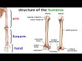
Understanding the Human Skeleton: A Detailed Overview of Bones and Structure
Explore the intricate structure of the human skeleton, comprising bones, cartilage, and more. Learn about axial and appendicular sections.
Comprehensive Overview of Neuroanatomy: High Yield Concepts for Exams
This video provides an in-depth discussion of high yield concepts in neuroanatomy, covering essential topics such as gray and white matter, brain vesicles, cranial nerves, and the structure of the spinal cord. Ideal for students preparing for exams, the content is structured to clarify complex concepts and enhance understanding.

Understanding Anatomical Position and Body Planes Explained
Learn the standard anatomical position and the major body planes—sagittal, coronal, and transverse—with easy memory tricks. This guide clarifies how body planes divide the body into sections and offers practical examples for better retention.
Comprehensive Guide to Body Cavities and Membranes Explained
This video simplifies the anatomy of body cavities and their membranes, covering the dorsal and ventral cavities, their subdivisions, and associated membranes. Learn key features of the cranial, spinal, thoracic, abdominal, and pelvic cavities with detailed explanations and visual aids.

Comprehensive Overview of Brain and Spinal Cord Functions
Explore the detailed anatomy and functions of the brain and spinal cord, including brain regions, lateralization, neural pathways, and imaging techniques. Understand key structures like the cerebral cortex, limbic system, and brainstem, plus the spinal cord's role in reflexes and communication.
Most Viewed Summaries

A Comprehensive Guide to Using Stable Diffusion Forge UI
Explore the Stable Diffusion Forge UI, customizable settings, models, and more to enhance your image generation experience.

Kolonyalismo at Imperyalismo: Ang Kasaysayan ng Pagsakop sa Pilipinas
Tuklasin ang kasaysayan ng kolonyalismo at imperyalismo sa Pilipinas sa pamamagitan ni Ferdinand Magellan.

Mastering Inpainting with Stable Diffusion: Fix Mistakes and Enhance Your Images
Learn to fix mistakes and enhance images with Stable Diffusion's inpainting features effectively.

Pamamaraan at Patakarang Kolonyal ng mga Espanyol sa Pilipinas
Tuklasin ang mga pamamaraan at patakaran ng mga Espanyol sa Pilipinas, at ang epekto nito sa mga Pilipino.

How to Install and Configure Forge: A New Stable Diffusion Web UI
Learn to install and configure the new Forge web UI for Stable Diffusion, with tips on models and settings.

If you found this summary useful, consider buying us a coffee. It would help us a lot!