Comprehensive AP Biology Unit 2 Review: Cell Structure & Function
Introduction to Cell Structure and Function
This AP Biology Unit 2 review covers the microscopic world of the cell, focusing on key organelles and their roles, membrane structure and permeability, and transport mechanisms essential for cellular life.
Key Organelles and Their Functions
- Ribosomes: Made of ribosomal RNA and protein, these non-membrane-bound structures synthesize proteins via translation, fundamental to all life forms.
- Endomembrane System: Includes rough and smooth endoplasmic reticulum (ER), Golgi complex, nuclear envelope, lysosomes, transport vesicles, and vacuoles. Rough ER aids protein synthesis; smooth ER synthesizes lipids and detoxifies; Golgi modifies and packages proteins. For a deeper understanding of this system, check out our Comprehensive Summary of Cell as the Unit of Life.
- Lysosomes: Contain hydrolytic enzymes to break down waste and damaged parts, also involved in apoptosis.
- Vacuoles: Storage units varying by cell type; central vacuole in plants stores water and nutrients, maintaining turgor pressure.
- Mitochondria: Double-membraned organelles where aerobic respiration occurs; inner membrane folds (cristae) increase ATP production. Learn more about the importance of these organelles in our Understanding Cell Organelles: A Quick Review and Ratings.
- Chloroplasts: Found in plants and algae, site of photosynthesis with thylakoid stacks converting light to chemical energy.
Cell Size and Surface Area to Volume Ratio
- Cells must maintain a high surface area to volume ratio for efficient nutrient uptake and waste removal.
- As cells grow, volume increases faster than surface area, limiting efficiency.
- Adaptations include membrane folds to increase surface area without increasing volume.
- This ratio also influences whole organism metabolism and heat regulation. For a comprehensive overview of these concepts, refer to our Understanding the Structure and Function of the Cell: A Comprehensive Overview.
Plasma Membrane Structure and Selective Permeability
- Composed of a phospholipid bilayer with hydrophilic heads and hydrophobic tails, creating a flexible, selective barrier.
- Embedded proteins facilitate transport, signaling, and structural support.
- The fluid mosaic model describes the dynamic and diverse membrane composition.
Membrane Transport Mechanisms
- Passive Transport: Includes diffusion, facilitated diffusion, and osmosis; moves substances down concentration gradients without energy.
- Facilitated Diffusion: Uses channel proteins for ions and aquaporins for water, enabling selective passage.
- Active Transport: Requires ATP to move substances against concentration gradients, exemplified by the sodium-potassium pump maintaining membrane potential. For more on transport mechanisms, see our article on Understanding Membrane Transport: Mechanisms and Importance.
- Bulk Transport: Endocytosis and exocytosis manage large molecule movement via vesicles.
Osmoregulation and Tonicity
- Water moves from hypotonic (low solute) to hypertonic (high solute) environments via osmosis.
- Cells regulate internal water and solute balance to prevent shrinkage or bursting.
- Water potential and solute potential calculations are critical for understanding these processes.
Cell Compartmentalization and Evolution
- Compartmentalization organizes cellular processes, increasing efficiency and surface area for reactions.
- Eukaryotic organelles like mitochondria and chloroplasts originated from endosymbiotic prokaryotes, supported by their own DNA and membranes.
- Prokaryotes lack membrane-bound organelles but have specialized internal regions.
Summary
Understanding cell structure and function is vital for grasping biological processes and excelling in AP Biology. This unit covers organelle roles, membrane dynamics, transport methods, and evolutionary insights, providing a foundation for further study.
Additional Resources
- Download the free AP Bio Ultimate Review Packet for study guides, practice questions, and video walkthroughs.
- Use the Ultimate Exam Slayer for focused exam preparation including practice tests and tips.
Prepare confidently for your quizzes, midterms, finals, and the AP Biology exam with these comprehensive resources.
Buckle up, we're about to shrink down and go full fantastic voyage into the microscopic world of the cell. From
power generating mitochondria to the selective fortress of the plasma membrane, every structure has a story.
And spoiler alert, it all matters for your AP bio score. So whether you're the nucleus of your study group or just
floating around like cytoplasm, this unit 2 recap has you covered. Hey, I'm Melanie King from the Absolute
Recap and I make AP Bio easier with podcasts, study guides, videos, the ultimate review packet, and the ultimate
exam slayer. This updated unit 2 review video all about cell structure and function follows the AP Biology course
and exam description released by the College Board for fall 2025. But it's more than just prep for your next quiz.
Think of it as your go-to tool for your midterm, your final, and of course, the AP Bio exam in May. Before we get
started, don't forget to download your free copy of the AP Bio Ultimate Review Packet. The links in the description.
It's packed with a video align study guide with an answer key, multiplechoice practice, skillbuilding worksheets with
video walkthroughs, and links to even more videos and podcasts. Not currently in AP Bio? No problem. These resources
are great for any high school or college level biology course, too. And if you're just here for a quick review, then check
out the Ultimate Exam Slayer. Focused 100% on exam prep. You'll get access to unit tests, fulllength practice tests,
one pages for every unit, math and graphing review sheets, and exclusive unit and exam tip videos. Okay, let's
zoom in. 2.1 cell structure and function. First up, ribosomes. These small but mighty structures are made of
two key ingredients. Ribosomal RNA, RRNA, and protein. Ribosomes are not membrane bound, which is an important
distinction. You'll find them floating freely in the cytoplasm or attached to the rough endopplasmic reticulum. More
on that in a bit. Why do they matter? Because ribosomes synthesize proteins based on instructions carried by
messenger RNA. This process is called translation and it's the central dogma of molecular biology. The fact that
ribosomes are found in all forms of life from bacteria to humans supports the theory of common ancestry. Every living
thing needs to make proteins and ribosomes make that possible. Next, let's look at the endommembrane system.
A group of membranebound organels that work together like a cellular assembly line to modify, package, and transport
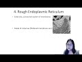
within the cell and beyond. Here's the breakdown. The endopplasmic reticulum, ER, comes in two varieties. Rough ER has
ribosomes attached. It helps compartmentalize the cell and plays a major role in protein synthesis and
processing. Smoothie R on the other hand has no ribosomes. It handles lipid synthesis and helps detoxify substances
in the cell. Think of it as the cellular version of a detox spa. The Golgi complex consists of flattened sacks that
fold, chemically modify, and package proteins for their final destination. This could be within the cell or out to
the extracellular environment. The system also includes the nuclear envelope, plasma membrane, losomes,
transport vesicles, and vacules, all working together to keep the traffic of materials flowing smoothly. Let's talk
about waste and storage because even cells need a cleanup crew. Losomes are membrane bound sacks filled with
hydraytic enzymes that break down damaged cell parts, waste, and even pathogens. They also play a key role in
apoptosis or programmed cell death, which is essential for development and disease prevention. Vacules are membrane
enclosed storage units with different functions depending on the type of cell. In plant cells, the central vacule is
huge and crucial. It stores water and nutrients and maintains trigger pressure, helping the plant stay
upright. In animal cells, vacules are smaller and more numerous, storing ions, nutrients, and waste products. Now for
some serious energy talk. The mitochondria. These double membraned organels are the site of aerobic
cellular respiration. The outer membrane is smooth but the inner membrane is highly folded into structures called
christe. This folding increases surface area allowing for more ATP production the energy currency of the cell. The
compartments created by these membranes allow different metabolic reactions to occur in an organized efficient way. So
more reactions equals more energy and more function. Last but not least chloroplasts. These are found only in
plants and photosynthetic algae and are the site of photosynthesis. Like mitochondria, chloroplast have a double
membrane and inside they contain stacks of membranes called philoids where light energy gets converted into chemical
energy glucose. Just like mitochondria, chloroplast create compartments to increase efficiency. Got all that? I
know there are a lot of organels to understand. I highly recommend trying the unit 2 practice sheet and video
inside the ultimate review packet to sharpen your organel identification skills by structure and function. 2.2.
Cell size. What does surface area and volume actually mean for cells? Surface area refers to the total area of the
cell's outer membrane, the interface between the inside of the cell and the environment. Volume is the amount of
space inside the cell. Basically, the cell's stuff or cytoplasm and organels. For cells to survive, the surface area
has to be big enough to support the needs of volume inside, like supplying nutrients and removing waste. Here's the
catch. As cells get bigger, its volume grows much faster than its surface area. This means the surface area to volume
ratio actually decreases, and that's a problem. A smaller ratio means less membrane surface to exchange materials
per unit of volume inside the cell. So, the cell can't get what it needs or get rid of waste efficiently. That's why
cells are small, easier to keep a high surface area to volume ratio and stay efficient. If cells get too big, they'd
starve internally or get poisoned by waste buildup because their membranes just can't keep up. Some cells and
organels solve this by having lots of folds or extensions in their membranes, increasing surface area without making
the structure huge. But the surface area to volume ratio doesn't just affect single cells, it affects whole
organisms, too. As animals get bigger, their surface area to volume ratio decreases, which changes how quickly
they gain or lose heat. Smaller animals lose heat quickly and have higher metabolic rates to stay warm, while
bigger animals hold heat longer and tend to have slower metabolisms. This explains why a tiny shrews heart beats
faster and it eats constantly, while an elephant's metabolism is much slower. Surface area and volume calculations
help biologists predict how cell size affects function and efficiency. All of the equations you need are on the
formula sheet. And if you need help understanding the math, check out the math review sheet in the ultimate exam
slayer. 2.3 plasma membrane. At the heart of the cell membrane is the phospholipid billayer, a double layer of
molecules that have split personalities. Each phospholipid has a hydrophilic head, meaning it loves water and
hydrophobic tails that avoid water at all costs. These molecules arrange themselves with their heads facing the
watery environments inside and outside the cell while the fatty acid tails huddle together in the middle away from
water. This setup forms a flexible but protective barrier that's selective about what crosses through. But the
membrane isn't just made of lipids. Scattered throughout are proteins. Some float, some anchor, and each one plays a
unique role. These proteins can be hydrophilic, hydrophobic or a combination of both depending on the
side chains of their amino acids. These proteins are involved in transport, signaling and maintaining structure. So
why is it called a fluid mosaic model? Fluid refers to the fact that the membrane components aren't locked in
place. They move and shift within the layer. Mosaic refers to the variety of components embedded in the membrane.
phospholipids, proteins, cholesterol, glyoproteins, and glyolippids forming a complex colorful patchwork. 2.4 Membrane
permeability. The plasma membrane's job is to separate the internal environment of the cell from the outside world, and
it does so with selective permeability. So, what actually makes it pass? Small non-polar molecules like nitrogen gas,
oxygen, and carbon dioxide slip right through, no problem. But larger polar molecules or ions, they need assistance.
And that's where embedded channels and transport proteins come in. Now, the plasma membrane isn't working alone.
Some cells have an extra layer of security. The cell wall found in bacteria, archa, fungi, some proise, and
plants. The cell wall provides a sturdy structural boundary. Importantly, it protects against osmotic lis. That
dangerous situation when too much water rushes in and the cell could literally burst. 2.5 Membrane transport. Selective
permeability is essential because it allows cells to create concentration gradients, differences in the amount of
a substance inside versus outside the cell. Without those gradients, cells couldn't power many of the processes
that keep life going. Now, let's talk about how molecules actually move across the selective barrier. The first method
is passive transport. Think of it like rolling a ball downhill. molecules naturally move from areas of high
concentration to areas of low concentration and the cell doesn't have to spend any energy. Diffusion,
facilitated diffusion, and osmosis all fall into this category. But sometimes cells need to move molecules against the
flow, like pushing a ball uphill. That's where active transport comes in. Active transport requires energy, usually in
the form of ATP, because it's moving substances from low concentration to high concentration, which doesn't happen
naturally. So what about molecules that are simply too big to squeeze through the membrane no matter what? That's when
cells rely on bulk transport, endoccytosis and exocytosis. In endoccytosis, the cell wraps its plasma
membrane around the material from the outside, creating little vesicles to pull it in. In exocytosis, it's the
opposite. Vesicles inside the cell fuse with the plasma membrane and release their contents out into the environment.
2.6 Facilitated diffusion. Facilitated diffusion is like a special entrance that helps certain molecules passively
get across the membrane. Think of it like a toll-free bridge. You're still going downhill, but you need a structure
to cross safely. For charged ions, the membrane is a total barrier without channel proteins. These ions rely on
proteins that act like tunnels carved straight through the lipid blayer. As they move, they can change the
electrical balance across the membrane. We call that polarization. If you've ever rubbed a balloon on your hair and
stuck it to the wall, you've seen static electricity in action. Cells use a similar trick with ions to create
charges across their membranes. Now, what about water? It may seem small, but moving lots of water at once isn't so
simple. That's why cells use aquaporns, protein channels dedicated to transporting huge amounts of water
quickly and efficiently. 2.7. Tenicity and osma regulation. When it comes to water movement in and out of cells, the
external environment makes all the difference. A solution can be hypotonic with less solute concentration,
hypertonic with greater solute concentration, or isotonic compared to the inside of the cell. The rule of
thumb here is simple. Water always moves from hypotonic to hypertonic regions. Or if you like to think of it in terms of
potential, water flows downhill from areas of high water potential to areas of low water potential. This process,
osmosis, is passive and doesn't require energy. But living organisms aren't just passive bystanders. They've got to
regulate water and solute balance to survive. That's where osmo regulation comes in. The body's way of making sure
cells don't shrivel up like raisins or burst like overfilled balloons. Osmo regulation helps maintain growth and
homeostasis by carefully controlling solute composition and water potential inside cells. The equations for water
potential and solute potential are on the equations and formula sheet provided on the exam. A higher solute
concentration means lower solute potential. Basically, the saltier it gets, the less water potential there is.
And calculated negative values for solute potential totally make sense because adding solutes makes water
potential drop. If you need more help with these calculations, you'll find it in the unit 2 practice sheet in the URP
and math practice sheet in the UEES. 2.8 Mechanisms of transport. When it comes to moving molecules and ions across the
cell membrane, sometimes simple diffusion isn't enough. And that's where active transport comes in. Without it,
larger charged molecules couldn't get where they need to go, and the whole system would stall. Membrane proteins
decide what gets transported, and they use energy from ATP to push molecules against their concentration gradient.
One of the most famous of these transport proteins is the sodium potassium pump. For every cycle, three
sodium ions are pushed out of the cell and two potassium ions are brought in. This uneven exchange creates and
maintains what we call a membrane potential, an electrical difference across the membrane. You can picture
this like charging a battery. The pump sets up an electrochemical gradient storing potential energy that the cell
can use later, just like your phone stores charge until you need it. In ATP, that's the currency funding the whole
operation. 2.9 Cell compartmentalization. If you tried to chop vegetables, boil pasta, and bake
cookies all on the same counter at the same time, things would get messy fast. But when you use separate cutting
boards, pots, and ovens, each task happens in its own space without interfering with the others. And that's
exactly what compartmentalization does in cells. It keeps reactions organized and efficient. Internal membranes also
solve another big problem, space. By folding and stretching out into complex shapes, they massively increase surface
area. More surface area means more places for enzymes to work and reactions to happen. Mitochondria, for example,
pack their inner membranes with folds of Christ, giving them the surface area needed to churn out lots of ATP. 2.10
origins of cell compartmentalization. When we look at the big differences between proaryotic and ukareotic cells,
one of the biggest gamech changers is the presence of membranebound organels. Ukareotic cells have these separate
compartments. Well, proarotes like bacteria generally do not. But here's the twist. Some of those ukareotic
organels actually started out as their own free-living proaryotic cells based on their own DNA, ribosomes, and double
membranes. The leading explanation is called the endo symbiotic theory. Imagine one cell swallowing another not
to digest it but to form a partnership. Over time, the engulfed procarot became a permanent resident evolving into
organels like mitochondria and chloroplast. Meanwhile, proarotic cells might not have these separate membrane
rooms, but they're not just a shapeless blob. They still have organized internal regions that carry out specialized
functions like the nucleoid where DNA hangs out or thyloin membranes and cyanobacteria which help capture light
energy. To recap, all living things are made of cells. Proarotic or ukareotic organels like the
nucleus, mitochondria, and chloroplast carry out essential cell functions. The endommembrane system coordinates protein
production and transport. The plasma membrane is selectively permeable thanks to phospholipids and proteins.
Diffusion, osmosis, and active transport move materials across membranes. Surface area to volume ratio affects how
efficient a cell can be. Compartmentalization lets ukarotic cells multitask efficiently. Mitochondria and
chloroplast have their own DNA, supporting the endo symbiotic theory. Overall, understanding cell structure
helps explain how life functions on every level. Need more support? Sign up for the ultimate review packet. The unit
2 free preview includes a study guide and practice multiple choice questions. And if you're ready to shift into full
exam mode, the ultimate exam slayer has practice tests, one pagers, and tips to help you dominate in May. All links are
in the description. Thanks for watching, and I'll see you next recap for unit 3, cellular energetics.
Key organelles include ribosomes, which synthesize proteins; the endomembrane system, which includes the rough and smooth endoplasmic reticulum (ER) for protein and lipid synthesis, and the Golgi complex for modifying and packaging proteins; lysosomes for breaking down waste; vacuoles for storage; mitochondria for aerobic respiration; and chloroplasts for photosynthesis in plants. Each organelle plays a crucial role in maintaining cellular function.
Cell size is critical because it influences the surface area to volume ratio, which affects nutrient uptake and waste removal. As cells grow, their volume increases faster than their surface area, potentially limiting efficiency. To adapt, cells may develop membrane folds to increase surface area without significantly increasing volume.
The plasma membrane is composed of a phospholipid bilayer with hydrophilic heads and hydrophobic tails, creating a flexible barrier. It contains embedded proteins that facilitate transport, signaling, and provide structural support, functioning as a selective barrier that regulates what enters and exits the cell.
Membrane transport mechanisms include passive transport (diffusion, facilitated diffusion, osmosis) that moves substances down concentration gradients without energy, and active transport that requires ATP to move substances against gradients, such as the sodium-potassium pump. Bulk transport methods like endocytosis and exocytosis manage the movement of large molecules.
Cells maintain water balance by regulating the movement of water from hypotonic (low solute) to hypertonic (high solute) environments via osmosis. This regulation prevents cells from shrinking or bursting, and involves calculations of water potential and solute potential to understand these processes.
Compartmentalization in eukaryotic cells organizes cellular processes, increasing efficiency and providing distinct environments for different reactions. This organization is supported by the presence of membrane-bound organelles, which are believed to have originated from endosymbiotic prokaryotes, enhancing the cell's functional capabilities.
For further study, you can download the free AP Bio Ultimate Review Packet, which includes study guides, practice questions, and video walkthroughs. Additionally, the Ultimate Exam Slayer offers focused exam preparation with practice tests and tips to help you prepare for quizzes, midterms, finals, and the AP Biology exam.
Heads up!
This summary and transcript were automatically generated using AI with the Free YouTube Transcript Summary Tool by LunaNotes.
Generate a summary for freeRelated Summaries
Understanding the Structure and Function of the Cell: A Comprehensive Overview
Explore the intricate details of cell structure and function in our comprehensive guide, including organelles and their roles.
Comprehensive Guide to Cell Biology: Free Revision Batch Lecture Summary
Explore the essentials of cell biology in our Free Revision Batch covering definitions, structures, and key discoveries.

Comprehensive AP Biology Study Plan and Review Guide for Exam Success
This guide offers a detailed, prioritized AP Biology study plan covering key units from molecular biology to ecology. Learn effective strategies like the stoplight method, understand crucial concepts such as cellular processes, genetics, evolution, and prepare with interactive resources for top exam performance.

Comprehensive Summary of Cell as the Unit of Life
This summary encapsulates the key points from a detailed lecture on the cell as the fundamental unit of life, covering topics such as cell theory, structure, function, and various organelles. It emphasizes the importance of understanding cellular components and their roles in biological processes.
Comprehensive Guide to Eukaryotic Cell Structure and Organelles
Explore the detailed functions and structures of key eukaryotic cell organelles including the nucleus, ribosomes, endoplasmic reticulum, Golgi apparatus, mitochondria, and chloroplasts. Understand protein synthesis, cellular transport, and energy production processes essential for cell function.
Most Viewed Summaries

A Comprehensive Guide to Using Stable Diffusion Forge UI
Explore the Stable Diffusion Forge UI, customizable settings, models, and more to enhance your image generation experience.

Kolonyalismo at Imperyalismo: Ang Kasaysayan ng Pagsakop sa Pilipinas
Tuklasin ang kasaysayan ng kolonyalismo at imperyalismo sa Pilipinas sa pamamagitan ni Ferdinand Magellan.

Mastering Inpainting with Stable Diffusion: Fix Mistakes and Enhance Your Images
Learn to fix mistakes and enhance images with Stable Diffusion's inpainting features effectively.

Pamamaraan at Patakarang Kolonyal ng mga Espanyol sa Pilipinas
Tuklasin ang mga pamamaraan at patakaran ng mga Espanyol sa Pilipinas, at ang epekto nito sa mga Pilipino.

How to Install and Configure Forge: A New Stable Diffusion Web UI
Learn to install and configure the new Forge web UI for Stable Diffusion, with tips on models and settings.

If you found this summary useful, consider buying us a coffee. It would help us a lot!