مقدمة
تبدأ الرؤية في العين، ولكن المعالجة العصبية للمعلومات البصرية تبدأ في شبكية العين التي تعد جزءًا من الجهاز العصبي المركزي.
مكونات العين ودورها في توجيه الضوء
- القرنية: طبقة شفافة في مقدمة العين تعكس وتقرب الضوء نحو الشبكية.
- العدسة: تعدل شكلها عبر العضلات الهدبية لضبط تركيز الضوء على شبكية العين، مع تناقص مرونتها مع التقدم في العمر، ما يؤثر على الرؤية القريبة.
- البؤبؤ والقزحية: ينظم حجم البؤبؤ كمية الضوء الواصلة للشبكية بحسب شدة الإضاءة الخارجية.
الشبكية ووظائف خلاياها العصبية
- تحتوي الشبكية على خمس أنواع رئيسية من الخلايا العصبية، وهي مرتبة في طبقات مميزة.
- تقع المستقبلات الضوئية في الطبقة الخلفية، وتشمل:
- العصي: حساسة جدًا للضوء، مسؤولة عن الرؤية في الإضاءة المنخفضة، لكنها لا تميز الألوان وتقدم دقة مكانية منخفضة.
- المخاريط: مسؤولة عن رؤية الألوان وتقدم دقة مكانية عالية، وتعمل في ظل ظروف إضاءة قوية.
- تحتوي الشبكية على مناطق متميزة مثل النقرة التي تهيمن عليها المخاريط وتوفر أعلى حدة بصرية.
معالجة الإشارات الضوئية في الشبكية
- عند امتصاص الفوتونات، تغير المستقبلات الضوئية إفراز الناقلات العصبية مما يؤثر على الخلايا الثنائية القطب ثم الخلايا العقدية.
- الخلايا الأفقية وعديمة الاستطالة تعدل وتُحسن الإشارات البصرية في مراحل معالجة مبكرة. لمزيد من التفاصيل حول الأنشطة العصبية وآليات نقل الإشارة، يمكن مراجعة شرح مفصل لجهد الفعل العصبي وآلية انتقال الإشارة في العصبونات.
نقل المعلومات من العين إلى الدماغ
- تحمل الخلايا العقدية الإشارات عبر العصب البصري، والذي يترك العين عبر القرص البصري (مما يخلق نقطة عمياء).
- في التصالب البصري، تتقاطع حوالي 60% من المحاور العصبية من الجانب الأنفي إلى الجانب المعاكس في الدماغ، مما يضمن معالجة المجال البصري الأيمن في الجانب الأيسر والعكس.
معالجة المعلومات البصرية في الدماغ
- بعد التصالب، تسمى المسارات العصبية الجهاز البصري وتصل إلى مناطق متعددة:
- Pretectum: مرتبط برد فعل انقباض الحدقة.
- النواة فوق التصالبية في ما تحت المهاد: تنسق الإيقاع اليومي بناءً على الضوء البيئي.
- الأكيمة العلوية: تنسق حركات الرأس والعين.
- الجزء الأكبر من الألياف تنتهي في النواة الركبية الجانبية للمهاد، التي ترسل الإشارات إلى القشرة البصرية الأولية عبر الإشعاعات البصرية.
القشرة البصرية الأولية والمناطق المرتبطة بها
- تقع القشرة البصرية الأولية (V1) حول التلم الكالكاريني، وهي مسؤولة عن تكوين الصورة المرئية الأولية.
- تستجيب خلاياها لخصائص مثل الحركة، الاتجاه، التباين والعمق.
- تتصل V1 بالمناطق البصرية الأخرى (V2, V3, V4, V5, V6)، التي تتخصص في جوانب معينة، مثل اكتشاف الحركة في V5.
- تسهم هذه المناطق في معالجة عالية المستوى، مثل التعرف على الأشكال وربط الصور بالخبرة السابقة. يمكن تعميق الفهم حول هذا الجانب من خلال قراءة Comprehensive Guide to Sensation and Perception in Psychology.
خاتمة
يقدم هذا المسار المعقد تحولاً متدرجًا للمعلومات من استقبال الضوء إلى فهم بصري متقدم في الدماغ، مما يمكننا من إدراك تفاصيل وألوان وحركة مشاهد العالم حولنا. لفهم أوسع للعلاقة بين بنية الدماغ والوظائف الشخصية المرتبطة، يمكن الاطلاع على Understanding the Brain: The Link Between Neuroanatomy and Personality.
Hi everyone, welcome to 10 minute neuroscience. In this installment, I’ll be covering the pathway of visual information through the brain starting with the eye and ending with the visual cortex and surrounding areas.
This’ll be a very general overview, and I’ll be focusing more on the pathway of visual information than on the processing of that information, but this should serve as a good introduction to the way visual information travels from the eye through the brain.
Of course, vision begins with the eye, but the neural aspect of vision really starts with the retina, which is the neural structure of the eye and is outlined in blue here. Other components of the eye, however, help to create a focused image on the retina.
This is accomplished to a large degree through the actions of the cornea and the lens. The cornea is a transparent layer at the front of the eye that lets light into the eye. It also bends, or refracts, those light rays to direct the light onto the retina.
The lens also helps to direct light on the retina. It has less refractive power than the cornea, but it has an advantage in that its shape can be modified by muscles in the eye called ciliary muscles, and changing the shape of
the lens can help to maintain focus for objects that are closer or farther away. As people get older, their lenses become less flexible and less capable of changing shape to focus on nearby objects; this is why people tend to need reading glasses when they get
older. The size of the pupil, the opening in the middle of the iris, which is the colored part of your eye, can be adjusted to regulate the amount of light that reaches the retina.
For example, there are muscles in the iris that cause the pupil to dilate (or open up more) in low-light situations, and constrict in an environment with higher levels of illumination since it doesn’t need to let in as much light in that type of environment.
The retina is a neural structure, and it’s actually considered part of the central nervous system. Its main function is to detect light and use that light to produce electrical and chemical
signals that the rest of the nervous system can understand. You can see here a close-up of a section of the retina, and although there are over 1,000 different types of neurons in the nervous system, there’s only 5 basic types of neurons
in the retina, and those neurons are situated in distinct layers. So, even though there are hundreds of millions of neurons in the retina, compared to the rest of the nervous system it’s relatively simple anatomically.
This has helped us to develop a better understanding of vision than of any other sensory system. Surprisingly, the layer of the retina that’s at the very back of the eye is the layer that contains photoreceptors, the cells responsible for converting light energy into electrochemical
signals, a process known as phototransduction. The location of the photoreceptors is surprising because it seems counterintuitive to have the light detecting cells at the very back of the retina, so light has to travel through
the eye and then through several layers of cells to reach the photoreceptors, but it’s thought that their location is strategic in the sense that they’re next to a layer of the retina called the pigment epithelium, and the cells of the pigment epithelium help
to maintain photoreceptor cells and keep them functioning properly. There are two main types of photoreceptor cells: rods and cones, which are named for their shape as you can see here.
These photoreceptor cells are the site where vision really begins; they each contain hundreds of disks that are capable of absorbing photons of light and absorbing photons causes the photoreceptors to change levels of neurotransmitter release in order to convey information about
a visual scene. Rods and cones have different functional specializations; they’re involved in distinct aspects of vision.
First off, cones enable us to see color, while rods don’t provide for color perception. Rods are also very sensitive to light and have low spatial resolution, meaning they’re not good at seeing details.
Cones, on the other hand, are not very sensitive to light and have high spatial resolution, so they provide us with higher visual acuity. In low light conditions, only rods are activated.
This makes it more difficult for us to make out details when there’s very little light due to the poor spatial resolution of rods, and it also means that in dim light we can’t perceive color.
When levels of illumination increase, eventually rods stop responding and fail to convey information. Essentially their high sensitivity to light causes them to become overstimulated, or saturated as its often called, in normal lighting situations like sunlight or even typical indoor lighting.
So in those conditions, cones are the dominant photoreceptor in determining how we see. Surprisingly, even though cones mediate perception in typical light situations, we have far more rods than cones in the retina.
There’s somewhere around 90 million rods and only about four and a half million cones. However, there is one part of the retina, an area called the fovea, where there are many more cones than rods.
In fact, at the very center of the fovea, which is called the foveola, there are no rods at all. Because of its high cone content the fovea is the part of our retina that has the capacity
for our highest acuity vision. This causes us to unconsciously move our eyes so that important visual information lands on our fovea, since this is the part of our eye most capable of discerning important details.
When photoreceptors absorb photons, it causes changes in the amount of neurotransmitters these cells release, and this affects the activity of the next layer of cells: bipolar cells.
Bipolar cells pass on signals about perceived light to the next layer of cells, which are called ganglion cells, and these cells will carry the visual information to the brain. We’ll talk more about that in a moment but I want to mention the other two major cell
types in the retina: horizontal cells and amacrine cells. Horizontal cells have dendrites that spread horizontally and make contact with multiple photoreceptor cells.
Horizontal cells modulate the function of photoreceptor cells to do things like enhance contrast and adapt to changes in lighting conditions, among other things. Amacrine cells make contact with bipolar cells, ganglion cells, and other amacrine cells,
and they also have a number of functions, but like horizontal cells they’re generally thought to be involved with refining the visual signal through their ability to modify the functions of other retinal cells.
So horizontal and amacrine cells are involved with very early processing of visual information. But most of the visual processing occurs in the brain, so the information from the retina has to be carried out of the eye and to the brain.
This is accomplished by the ganglion cells, whose axons leave the eye in a bundle at a region called the optic disc. Because the optic disc is the area where the ganglion cells leave the eye, and essentially
they need a place where they can exit the eye, there are no photoreceptors there. So, this creates a small region where we don’t receive any visual information, or a blind spot.
Amazingly, we don’t notice this blind spot in our visual scene because the brain fills it in with information from other photoreceptors. If you find it hard to believe that you’ve got a blind spot always present in your field
of vision, click on this link above for a quick and simple experiment that proves the existence of your blind spot. The ganglion cells leaving the eye form the optic nerve, which is one of our cranial nerves.
The optic nerve extends back to this region just below the hypothalamus called the optic chiasm. At the optic chiasm, about 60% of the axons from the optic nerve cross over to the other
side of the brain, while the rest stay on the side they originated on. The fibers that are coming from the nasal part of the retina—the part closer to the nose—cross over to the other side, or decussate, while the fibers from the temporal part of
the retina—the part closer to the temple–do not cross over. The result of this is that all of the information from the right visual field ends up traveling to the left side of the brain, and vice versa.
After the optic chiasm, the visual fibers are no longer called the optic nerve—they’re now called the optic tract. The optic tracts extend to multiple areas.
For example, some of the fibers go to an area in the brainstem called the pretectum, which is involved in a number of visual functions such as the pupillary light reflex, which causes your pupils to constrict when there’s greater illumination in your environment.
Other fibers go to a region of the hypothalamus called the suprachiasmatic nucleus; this nucleus helps to maintain circadian or daily rhythms and uses information about light in the environment to help to do that.
And still other fibers go to the superior colliculus, a region in the brainstem that among other things helps to coordinate head and eye movements to focus on objects of interest in the visual field.
Most of the fibers in the optic tracts, however, end in a nucleus in the thalamus called the lateral geniculate nucleus, there’s one of these on each side of the brain here. Here, the optic tracts synapse on neurons that leave the lateral geniculate nucleus
and extend toward the back of the brain as bundles of fibers called the optic radiations. The optic radiations travel back to a region of cortex that surrounds a fissure called the calcarine sulcus.
This small area of cortex that surrounds the calcarine sulcus is called the primary visual cortex, or V1, it's represented by this striped area here. There’s a collection of myelinated fibers that forms a white stripe here that can be
seen with the naked eye in anatomical brain sections of this region and because of this striation or stripe, sometimes the primary visual cortex is called the striate cortex. The primary visual cortex helps to make a visual image out of the information that has
been received by the retina. To do that, there are neurons in the primary visual cortex that are activated preferentially by different characteristics of a visual stimulus, such as orientation, movement, contrast, depth,
etc. The primary visual cortex also communicates with a multitude of other visual areas that surround it, including visual area 2, or V2, visual area 3 or V3, V4, V5, and V6.
Neurons in these other areas seem to be specialized to some degree for detecting specific aspects of a visual scene. Neurons in V5, for example, seem to be specialized for detecting movement.
These additional visual areas are recruited by V1 and they also begin to recruit other areas of the brain to accomplish higher-level processing of visual information. This enables our brain to move from identifying the basic aspects of a visual scene, such
as shape and contrast, to more complex tasks like object recognition, which can provide the image in our brain with meaning based on previous experience. So we just scratched the surface there but I hope that gave you a sense of the major
pathway that visual information travels in the brain. Thanks for watching.
تبدأ الرؤية بدخول الضوء عبر القرنية والعدسة التي تركزه على شبكية العين، حيث تحتوي على المستقبلات الضوئية (العصي والمخاريط) التي تُحوّل الضوء إلى إشارات عصبية تبدأ معالجة المعلومات البصرية في الجهاز العصبي المركزي.
الخلايا العصوية حساسة جداً للضوء وتعمل في الإضاءة المنخفضة لكنها لا تميز الألوان وتقدم دقة مكانية منخفضة، بينما المخاريط مسؤولة عن رؤية الألوان وتعمل في ظروف إضاءة قوية مع تقديم دقة مكانية عالية، خصوصاً في منطقة النقرة.
تحمل الخلايا العقدية الإشارات العصبية عبر العصب البصري الذي يخرج من العين، وتتقاطع نسبة من هذه المحاور في التصالب البصري لتصل إلى الجانب المعاكس من الدماغ، مما يضمن معالجة المجال البصري الأيمن في الجانب الأيسر والعكس.
تصل الإشارات إلى مناطق متعددة مثل Pretectum المسؤول عن ضبط حدقة العين، والنواة فوق التصالبية التي تنسق الإيقاع اليومي، والأكيمة العلوية التي تنسق حركات الرأس والعين، بالإضافة إلى النواة الركبية الجانبية التي ترسل المعلومات إلى القشرة البصرية الأولية.
تقع V1 حول التلم الكالكاريني وتكوّن الصورة المرئية الأولية، وتستجيب لخصائص مثل الحركة، الاتجاه، التباين والعمق، وترسل المعلومات إلى مناطق بصرية أخرى تخصصية تعالج الجوانب المعقدة مثل اكتشاف الحركة والتعرف على الأشكال.
تُعدل العدسة شكلها عبر العضلات الهدبية لضبط تركيز الضوء على الشبكية، مما يسمح برؤية واضحة للأجسام القريبة والبعيدة، ومع التقدم في العمر تقل مرونة العدسة مما يؤثر سلباً على القدرة على الرؤية القريبة.
النقطة العمياء تحدث عند القرص البصري حيث يخرج العصب البصري من العين ولا توجد مستقبلات ضوئية هناك، لهذا الجزء لا يستطيع رصد أي ضوء أو صورة مما يخلق نقطة عمياء في مجال الرؤية.
Keep this summary
Save it to LunaNotes and it becomes a real note in your library — editable, searchable, and ready to turn into flashcards or a diagram. Free to start.
Save to LunaNotesOr summarise for another video.
This summary and transcript were automatically generated using AI with the Free YouTube Transcript Summary Tool by LunaNotes.
Related summaries
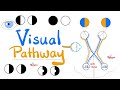
Detailed Visual Pathway Anatomy and Associated Visual Deficits Explained
Explore the comprehensive anatomy of the visual pathway, from the retina through the optic nerve to the visual cortex, including detailed discussion on fiber crossings, optic radiations, and clinical deficits such as hemianopia and quadrant anopsia. Understand how lesions at different points affect visual fields and learn key mnemonics for retention.

شرح مفصل لجهد الفعل العصبي وآلية انتقال الإشارة في العصبونات
يشرح هذا الفيديو كيفية تكوين العصبونات وأجزاءها الرئيسية، وآلية استقبالها ونقلها للإشارات العصبية عبر جهد الفعل. يتناول الفيديو دور القنوات الشاردية المبوبة بالفولتاج والميالين في تسريع انتقال الإشارة العصبية.
Anatomie du cerveau antérieur : cortex, ganglions de la base et diencéphale
Ce module de cours explore en détail le cerveau antérieur (prosencéphale), en décomposant ses deux grandes structures : le télencéphale (cortex cérébral, matière blanche et noyaux gris centraux) et le diencéphale (thalamus et hypothalamus). Vous découvrirez l'organisation des lobes, les circonvolutions, les aires de Brodmann, les fonctions motrices et sensorielles, ainsi que le rôle crucial des ganglions de la base dans le contrôle du mouvement, avec une mention de pathologies comme la maladie de Parkinson.

Comprehensive Guide to Sensation and Perception in Psychology
Explore the fundamentals of sensation and perception, including sensory processes, visual and auditory systems, and psychological interpretations. This summary highlights key concepts such as sensory receptors, thresholds, Gestalt principles, and the interplay between bottom-up and top-down processing.

Introduction to Neuroanatomy: Central and Peripheral Nervous Systems Explained
This comprehensive overview simplifies basic neuroanatomy concepts, exploring the central and peripheral nervous systems, sensory and motor functions, gray and white matter, and key brain structures. Learn how sensory information is processed, motor responses generated, and how different neural pathways and cellular components contribute to nervous system function.
Most viewed summaries

A Comprehensive Guide to Using Stable Diffusion Forge UI
Explore the Stable Diffusion Forge UI, customizable settings, models, and more to enhance your image generation experience.

Kolonyalismo at Imperyalismo: Ang Kasaysayan ng Pagsakop sa Pilipinas
Tuklasin ang kasaysayan ng kolonyalismo at imperyalismo sa Pilipinas sa pamamagitan ni Ferdinand Magellan.

Mastering Inpainting with Stable Diffusion: Fix Mistakes and Enhance Your Images
Learn to fix mistakes and enhance images with Stable Diffusion's inpainting features effectively.

Pamamaraan at Patakarang Kolonyal ng mga Espanyol sa Pilipinas
Tuklasin ang mga pamamaraan at patakaran ng mga Espanyol sa Pilipinas, at ang epekto nito sa mga Pilipino.

How to Install and Configure Forge: A New Stable Diffusion Web UI
Learn to install and configure the new Forge web UI for Stable Diffusion, with tips on models and settings.
Found this summary useful?
Take it with you. One click puts it in your own LunaNotes library.
Save to LunaNotes